. 2019 Aug 1;76(8):915-924.
doi: 10.1001/jamaneurol.2019.1424.

Association of Amyloid and Tau With Cognition in Preclinical Alzheimer Disease: A Longitudinal Study

Affiliations

Association of Amyloid and Tau With Cognition in Preclinical Alzheimer Disease: A Longitudinal Study

Bernard J Hanseeuw et al. JAMA Neurol. .

Erratum in

  • Error in Figure 3.
    [No authors listed] [No authors listed] JAMA Neurol. 2019 Aug 1;76(8):986. doi: 10.1001/jamaneurol.2019.2144. JAMA Neurol. 2019. PMID: 31403691 Free PMC article. No abstract available.

Abstract

Importance: Positron emission tomography (PET) imaging now allows in vivo visualization of both neuropathologic hallmarks of Alzheimer disease (AD): amyloid-β (Aβ) plaques and tau neurofibrillary tangles. Observing their progressive accumulation in the brains of clinically normal older adults is critically important to understand the pathophysiologic cascade leading to AD and to inform the choice of outcome measures in prevention trials.

Objective: To assess the associations among Aβ, tau, and cognition, measured during different observation periods for 7 years.

Design, setting, and participants: Prospective cohort study conducted between 2010 and 2017 at the Harvard Aging Brain Study, Boston, Massachusetts. The study enrolled 279 clinically normal participants. An additional 90 individuals were approached but declined the study or did not meet the inclusion criteria. In this report, we analyzed data from 60 participants who had multiple Aβ and tau PET observations available on October 31, 2017.

Main outcomes and measures: A median of 3 Pittsburgh compound B-PET (Aβ, 2010-2017) and 2 flortaucipir-PET (tau, 2013-2017) images were collected. We used initial PET and slope data, assessing the rates of change in Aβ and tau, to measure cognitive changes. Cognition was evaluated annually using the Preclinical Alzheimer Cognitive Composite (2010-2017). Annual consensus meetings evaluated progression to mild cognitive impairment.

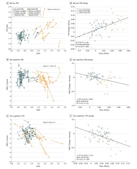
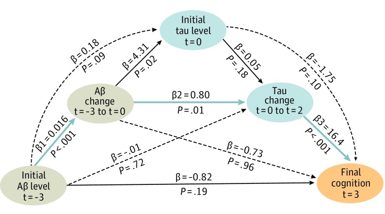
Results: Of the 60 participants, 35 were women (58%) and 25 were men (42%); median age at inclusion was 73 years (range, 65-85 years). Seventeen participants (28%) exhibited an initial high Aβ burden. An antecedent rise in Aβ was associated with subsequent changes in tau (1.07 flortaucipir standardized uptake value ratios [SUVr]/PiB-SUVr; 95% CI, 0.13-3.46; P = .02). Tau changes were associated with cognitive changes (-3.28 z scores/SUVR; 95% CI, -6.67 to -0.91; P = .001), covarying baseline Aβ and tau. Tau changes were greater in the participants who progressed to mild cognitive impairment (n = 6) than in those who did not (n = 11; 0.05 SUVr per year; 95% CI, 0.03-0.07; P = .001). A serial mediation model demonstrated that the association between initial Aβ and final cognition, measured 7 years later, was mediated by successive changes in Aβ and tau.

Conclusions and relevance: We identified sequential changes in normal older adults, from Aβ to tau to cognition, after which the participants with high Aβ with greater tau increase met clinical criteria for mild cognitive impairment. These findings highlight the importance of repeated tau-PET observations to track disease progression and the importance of repeated amyloid-PET observations to detect the earliest AD pathologic changes.


Conflict of interest statement

Conflict of Interest Disclosures: Dr Hanseeuw reported grants from the Belgian National Fund for Scientific Research and the Belgian Foundation for Alzheimer Research during the conduct of the study and personal fees from GE Healthcare outside the submitted work. Dr Jacobs reported funding from the European Union's Horizon 2020 Research and Innovation Programme under the Marie Sklodowska-Curie Grant agreement (IF-2015-GF, 706714). Dr Becker reported grants from the National Institutes of Health during the conduct of the study. Dr Quiroz reported grants from the National Institutes of Health and the National Institute on Aging during the conduct of the study. Dr Hedden reported grants from the National Institutes of Health during the conduct of the study. Dr Rentz reported other support from Eli Lilly, Neurotrack, and Biogen outside the submitted work. Dr Sperling reported grants from Janssen during the conduct of the study and personal fees from AC Immune, Biogen, and Roche outside the submitted work. Dr Johnson reported grants from the National Institutes of Health; personal fees from Biogen, Lilly/Avid, Merck, Novartis, Takeda, Roche/Genentech, and Janssen; and grants from Alzheimer’s Association and from Alzheimer’s Drug Discovery Foundation during the conduct of the study. No other disclosures were reported.

Figures

Figure 1.
Figure 1.. Research Design and Serial Tau–Positron Emission Tomography (PET) Surface Images of an Illustrative Participant
A, Baseline of this study was defined as the time of baseline FTP imaging (t = 0; where t indicates time in years from baseline). Change in flortaucipir (FTP) was evaluated between t = 0 and t = 2. Pittsburgh compound B (PiB) and cognitive data have been evaluated both between t = −3 and t = 0 and between t = 0 and t = 3. Parentheses mean that less than half the sample was observed. The brain images are of an illustrative participant with high PiB at baseline FTP-PET (ε4 noncarrier). Global Clinical Dementia Rating remained stable at 0 during the follow-up, but Preclinical Alzheimer Cognitive Composite z scores declined from 0.01 (t = 0) to −0.88 (t = 3). Note the progressive extension of FTP-PET signal from left entorhinal cortex to left temporal neocortex, posterior cingulate, and to the homologous regions in the right hemisphere. The FTP-PET images use a threshold set at standardized uptake value ratios (SUVr) of 1.05, with cerebral white matter as reference and partial volume correction.
Figure 2.
Figure 2.. Longitudinal Associations Between Amyloid-β (Aβ), Tau, and Cognition, Observed Contemporaneously
A-C, Spaghetti plots showing the unadjusted positron emission tomography (PET) standardized uptake value ratios (SUVr) and Preclinical Alzheimer Cognitive Composite (PACC) scores at the initial t = −3 (n = 50; where t indicates time in years from baseline), baseline t = 0 (n = 60), and follow-up t = 2 (N = 60) observations. All MCI progressors had high Pittsburgh compound B (PiB) signal; they were not different than other participants with high PiB at baseline (similar age, PACC, PiB, and FTP), and their PiB change was not particularly fast (B, vertical orange lines ending with a star). However, they had fast FTP and PACC changes (C plot, oblique orange lines). D-F, PiB, FTP, and PACC slope data observed simultaneously are plotted against each other. All associations are significant, although the PiB-PACC longitudinal association is weaker than the PiB-FTP or the FTP-PACC association (Table 2; model 7), probably reflecting that PiB and PACC changes are more distant in time than PiB and FTP or FTP and PACC changes.
Figure 3.
Figure 3.. Overview of Sequential Associations Between Amyloid-β (Aβ), Tau, and Cognition
Diagram of mediation model pathways relating Aβ, tau, and cognition. Each observation was measured at different, successive times. The mediation highlighted in blue (indirect effect: −0.21; 95% CI, −0.55 to −0.06; P = .06) accounts for 20% of the direct effect between initial Pittsburgh compound B (PiB) and final Preclinical Alzheimer Cognitive Composite (PACC) t = 3, where t indicates time in years from baseline. Altogether, the pathways explain 45% of the direct effect. Black dotted lines illustrate alternative pathways that were not significant. This serial mediation supports a temporal sequence of phenomena in preclinical Alzheimer disease. It is consistent with Table 2, models 1, 4, and 7. It is associated with final PACC t = 3 (not PACC slope as in model 7) to dissociate the time of the outcome measure from the time of the predictors measure. Sixty participants were included in this analysis, using baseline PiB t = 0 instead of initial PiB t = −3 for the 10 participants missing the initial PiB observation. Highly similar results were obtained when excluding these 10 participants.

Comment in

References

    1. McKhann GM, Knopman DS, Chertkow H, et al. . The diagnosis of dementia due to Alzheimer’s disease: recommendations from the National Institute on Aging-Alzheimer’s Association workgroups on diagnostic guidelines for Alzheimer’s disease. Alzheimers Dement. 2011;7(3):263-269. doi:10.1016/j.jalz.2011.03.005 - DOI - PMC - PubMed
    1. Hyman BT, Phelps CH, Beach TG, et al. . National Institute on Aging-Alzheimer’s Association guidelines for the neuropathologic assessment of Alzheimer’s disease. Alzheimers Dement. 2012;8(1):1-13. doi:10.1016/j.jalz.2011.10.007 - DOI - PMC - PubMed
    1. Price JL, McKeel DW Jr, Buckles VD, et al. . Neuropathology of nondemented aging: presumptive evidence for preclinical Alzheimer disease. Neurobiol Aging. 2009;30(7):1026-1036. doi:10.1016/j.neurobiolaging.2009.04.002 - DOI - PMC - PubMed
    1. Nelson PT, Alafuzoff I, Bigio EH, et al. . Correlation of Alzheimer disease neuropathologic changes with cognitive status: a review of the literature. J Neuropathol Exp Neurol. 2012;71(5):362-381. doi:10.1097/NEN.0b013e31825018f7 - DOI - PMC - PubMed
    1. Mintun MA, Larossa GN, Sheline YI, et al. . [11C]PIB in a nondemented population: potential antecedent marker of Alzheimer disease. Neurology. 2006;67(3):446-452. doi:10.1212/01.wnl.0000228230.26044.a4 - DOI - PubMed