

Targeted quantitative proteomic analysis of drug metabolizing enzymes and transporters by nano LC-MS/MS in the sandwich cultured human hepatocyte model
- PMID: 31158457
- PMCID: PMC6701468
- DOI: 10.1016/j.vascn.2019.106590
Targeted quantitative proteomic analysis of drug metabolizing enzymes and transporters by nano LC-MS/MS in the sandwich cultured human hepatocyte model
Abstract
Introduction: Sandwich-cultured human hepatocytes (SCHHs) are the most common in vitro hepatocyte model used for studying hepatic drug disposition and hepatotoxicity. Targeted quantification of key DME and transporter protein expression is useful for in vitro-in vivo extrapolation of drug and xenobiotic clearance and developing corresponding PBPK models. However, established methods for comprehensive quantification of drug metabolizing enzyme (DMEs) and transporter expression in SCHHs are lacking. In this study, a targeted quantitative proteomic isotope dilution nanoLC-MS/MS method developed in our laboratory was adapted to quantify a panel of phase I & II DMEs and transporter proteins in SCHHs under basal and induced conditions.
Methods: SCHHs were treated with known inducers of DMEs (Rifampin: PXR activator, CITCO: CAR activator) and transporters (CDCA: FXR activator) or with vehicle control (DMSO) for 72 h. Membrane protein was isolated from the SCHHs using a membrane extraction kit and 30 μg membrane protein was digested with trypsin. The resulting peptides were analyzed by isotope dilution nanoLC-MS/MS to quantify the DMEs and transporters.
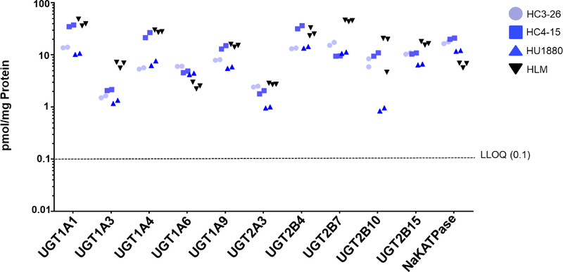
Results: Using the method, we could quantify fourteen phase I and ten phase II DMEs, and twelve uptake/efflux transporters, under basal and induced conditions in the SCHHs. Analysis showed donor to donor variation in basal protein levels of CYP450s, UGTs and transporters, and that basal protein expression of CYP450s and UGTs was higher than that of transporters. In addition, induction of key proteins in response to rifampin, CITCO and CDCA was observed.
Discussion: We have successfully quantified protein abundance of multiple phase I and II DMEs and uptake and efflux transporters in SCHHs using a method previously developed in our laboratory. Our method is sufficiently sensitive to quantify inter-donor differences in protein concentrations at the basal level as well as changes in protein expression in response to endogenous and exogenous stimuli.
Keywords: Drug metabolizing enzymes; Hepatocytes; Proteomics; Quantification; SCHH model; Targeted; Transporters.
Copyright © 2019 Elsevier Inc. All rights reserved.
Conflict of interest statement
Conflict of interest
The authors declared no competing financial interest.
Figures


References
-
- Badee J, Achour B, Rostami-Hodjegan A, & Galetin A (2015). Meta-Analysis of Expression of Hepatic Organic Anion-Transporting Polypeptide (OATP) Transporters in Cellular Systems Relative to Human Liver Tissue. Drug Metabolism and Disposition, 43, 424–432. - PubMed
-
- Bi YA, Kimoto E, Sevidal S, Jones HM, Barton HA, Kempshall S, Whalen KM, Zhang H, Ji CJ, Fenner KS, El-Kattan AF, & Lai YR (2012). In Vitro Evaluation of Hepatic Transporter-Mediated Clinical Drug-Drug Interactions: Hepatocyte Model Optimization and Retrospective Investigation. Drug Metabolism and Disposition, 40, 1085–1092. - PubMed
-
- Birdwell KA, Decker B, Barbarino JM, Peterson JF, Stein CM, Sadee W, Wang D, Vinks AA, He Y, Swen JJ, Leeder JS, van Schaik R, Thummel KE, Klein TE, Caudle KE, & MacPhee IA (2015). Clinical Pharmacogenetics Implementation Consortium (CPIC) Guidelines for CYP3A5 Genotype and Tacrolimus Dosing. Clin Pharmacol Ther, 98, 19–24. - PMC - PubMed
-
- Brouwer KLR, Keppler D, Hoffmaster KA, Bow DAJ, Cheng Y, Lai Y, Palm JE, Stieger B, Evers R, & Consortium IT (2013). In Vitro Methods to Support Transporter Evaluation in Drug Discovery and Development. Clinical Pharmacology & Therapeutics, 94, 95–112. - PubMed
MeSH terms
Substances
Grants and funding
LinkOut - more resources
Full Text Sources
