. 2019 Sep 1:177:386-400.
doi: 10.1016/j.ejmech.2019.05.068. Epub 2019 May 25.

Enhanced tumor retention of NTSR1-targeted agents by employing a hydrophilic cysteine cathepsin inhibitor

Affiliations

Enhanced tumor retention of NTSR1-targeted agents by employing a hydrophilic cysteine cathepsin inhibitor

Wei Fan et al. Eur J Med Chem. .

Abstract

We explored the approach of using an analog of E-64, a well-known and hydrophilic cysteine cathepsin (CC) inhibitor, as a potent cysteine cathepsin-trapping agent (CCTA) to improve the tumor retention of low-molecular-weight, receptor-targeted radiopharmaceuticals. The synthesized hydrophilic CCTA-incorporated, NTSR1-targeted agents demonstrated a substantial increase in cellular retention upon uptake into the NTRS1-positive HT-29 human colon cancer cell line. Similarly, biodistribution studies using HT-29 xenograft mice revealed a significant and substantial increase in tumor retention for the CCTA-incorporated, NTSR1-targeted agent. The intracellular trapping mechanism of the CCTA-incorporated agents by macromolecular adduct formation was confirmed using multiple in vitro and in vivo techniques. Furthermore, utilization of the more hydrophilic CCTA greatly increased the hydrophilicity of the resulting NTSR1-targeted constructs leading to substantial decreases in most non-target tissues in contrast to our previously reported dipeptidyl acyloxymethyl ketone (AOMK) constructs. This work further confirms that the CCTA trapping approach can make significant improvements in the clinical potential of NTSR1-and other receptor-targeted radiopharmaceuticals.

Keywords: Cysteine cathepsin inhibitor; E−64 analogue; NTSR1; Radiopharmaceutical; Trapping agent; Tumor retention.


Figures

Fig. 1.
Fig. 1.
Structures of the cysteine cathepsin inhibitors and peptidic conjugates studied in our previous and present work
Fig. 2.
Fig. 2.
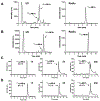
The RP-HPLC profiles of radiolabeling NE1a (A) and NE1b (B) by 177LuCl3. The stability and purity of 177Lu-NE1a (C) and 177Lu-NE1a (D) in human serum at 37 °C at various time points.
Fig. 3.
Fig. 3.
The hyperbolic regression of pseudo-first-order rate constants (kobs) (t = 0-60 min) by CatB (A) and CatL(B) versus the concentrations of NE1a ([C]). The substrates for CatB and CatL were Z-Arg-Arg-AMC and Z-Phe-Arg-AMC, respectively. The substrate concentration was from 1 to 500 nM. The Ki and ki/Ki determined from kobs = ki[C]/(Ki + [C]). (C) A dose-response curve showing the inhibition of NTSR1 receptor binding of 177Lu-N1 by NE1a and NE1b. Values are means ± SD (n = 3).
Fig. 4.
Fig. 4.
The internalized and surface-bound 177Lu-NE1a (A) and 177Lu-NE1b (B) by HT-29 cells. (C) Efflux profiles of the internalized 177Lu-NE1a and 177Lu-NE1b over 24 h in HT-29 cells. Values are means ± SD (n = 3).
Fig. 5.
Fig. 5.
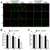
(A) Representative confocal microscopy images of the efflux of natEu labeled NE1a and NE1b (red) form HT-29 cells. Cell endolysosomal compartments were stained with Lysotracker (green). Scale bar = 50 μm. (B) Time-dependent fluorescence intensity of natEu per cell as quantified from the confocal images. (C) Co-localization efficiencies of natEu (red) overlapping with Lysotracker® (green). All the analysis was performed in 6 random images and were presented as mean ± SD. **p < 0.01, ***p < 0.001, NS = not significant.
Fig. 6.
Fig. 6.
The autoradiography of the SDS-PAGE showing the CatB and CatL binding with the 177Lu-NE1a and 177Lu-NE1b in the presence or absence of the cysteine proteases inhibitor E-64 (A) and the NTSR1 ligand N1 (B). (C). Autoradiographic image of an SDS-PAGE gel examining the time-dependent retention of CC-conjugate adducts in HT-29 cells after pre-incubation with 177Lu-NE1a for 4 h.
Fig. 7.
Fig. 7.
(A) The autoradiography of SDS-PAGE of the tumor, liver, and kidney samples at 72 h p.i. of 177Lu-NE1a and 177Lu-NE1b in mice. The radioconjugates were injected to the mice at a dose of 800 μCi (29.6 MBq) /mouse. (B) Percentage of the macromolecules associated radioactivity (Mw > 10 kDa) in tumor, liver, and kidney samples at 72 h after administration of the radioconjugates (n=3). *p < 0.05, **p < 0.01, ***p < 0.001, NS = not significant.
Scheme 1.
Scheme 1.
Synthetic Procedures for 1a and 1ba aReagents and conditions: (a) (i) EDCI, NHS, DMF, 0°C, 2-3h (ii) 2-azidoethanamine, DIEA,DMF, r.t., overnight; (b) (i) 20% piperidine in DMF, r.t., 30min; (ii) COMU, Fmoc-Leu-OH, DIEA, DMF, r.t., 2h; (c) (i) 20% piperidine in DMF, r.t., 30min; (ii) COMU, (+/−)-trans-oxirane-2,3-dicarboxylic acid, DMF, DIEA, r.t., overnight; (c) (i) 20% piperidine in DMF, r.t., 30min; succinic anhydride, TEA, DMF, 50°C, overnight; (e) 90% TFA in DCM, r.t., 2h.
Scheme 2.
Scheme 2.
Synthetic Procedures for NE1a and NE1ba aReagents and conditions: (a) CuSO4, ascorbic acid, TEA, H2O:n-Butanol:DMF (1:1:2), r.t., 1h; (b) (i) DOTA-NHS, DIEA, DMF, r.t., overnight; (ii) 90% TFA in DCM, r.t., 3h.

References

    1. Gambhir SS, Molecular imaging of cancer with positron emission tomography, Nat. Rev. Cancer 2 (2002) 683–693. - PubMed
    1. Okarvi S, Peptide-based radiopharmaceuticals: Future tools for diagnostic imaging of cancers and other diseases, Med. Res. Rev 24 (2004) 357–397. - PubMed
    1. Srinivasarao M, Galliford CV, Low PS, Principles in the design of ligand-targeted cancer therapeutics and imaging agents, Nat. Rev. Drug Discov 14 (2015) 203–219. - PubMed
    1. Okarvi S, Peptide-based radiopharmaceuticals and cytotoxic conjugates: potential tools against cancer, Cancer Treat. Rev 34 (2008) 13–26. - PubMed
    1. Fani M, Maecke H, Okarvi S, Radiolabeled peptides: valuable tools for the detection and treatment of cancer, Theranostics 2 (2012) 481–501. - PMC - PubMed

MeSH terms

LinkOut - more resources