. 2019 May 31;11(6):1241.
doi: 10.3390/nu11061241.

Priming of Hypothalamic Ghrelin Signaling and Microglia Activation Exacerbate Feeding in Rats' Offspring Following Maternal Overnutrition

Affiliations

Priming of Hypothalamic Ghrelin Signaling and Microglia Activation Exacerbate Feeding in Rats' Offspring Following Maternal Overnutrition

Roger Maldonado-Ruiz et al. Nutrients. .

Abstract

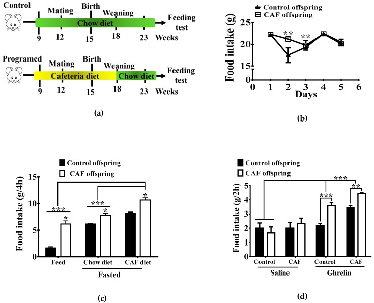
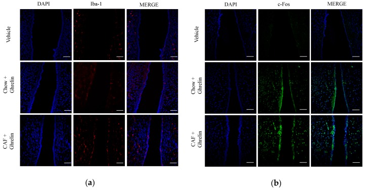
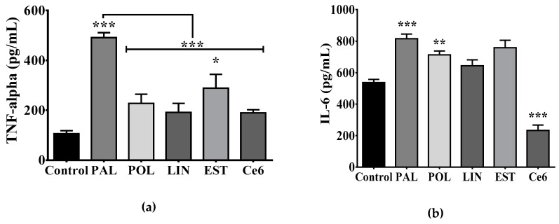
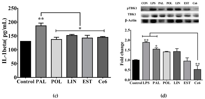
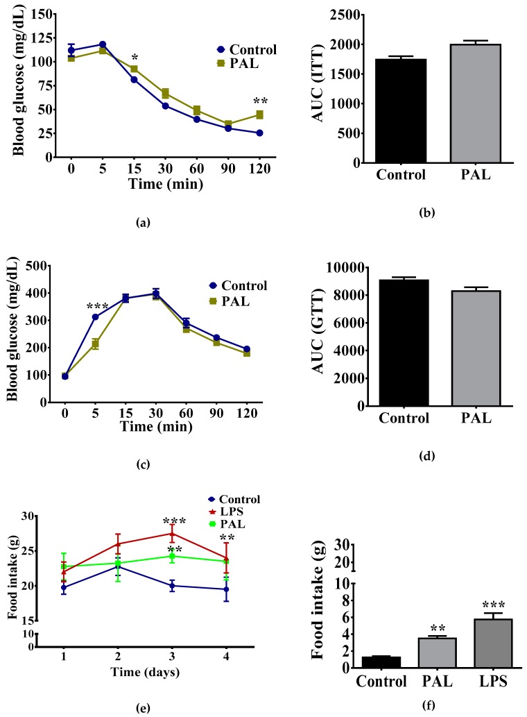
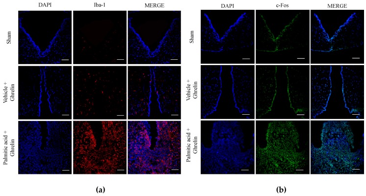
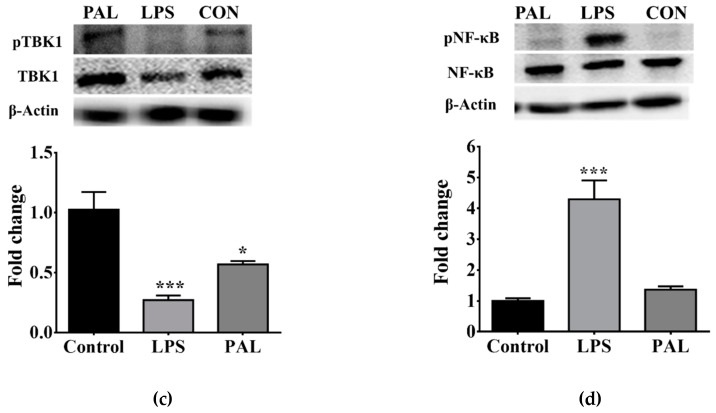
Maternal overnutrition during pregnancy leads to metabolic alterations, including obesity, hyperphagia, and inflammation in the offspring. Nutritional priming of central inflammation and its role in ghrelin sensitivity during fed and fasted states have not been analyzed. The current study aims to identify the effect of maternal programming on microglia activation and ghrelin-induced activation of hypothalamic neurons leading to food intake response. We employed a nutritional programming model exposing female Wistar rats to a cafeteria diet (CAF) from pre-pregnancy to weaning. Food intake in male offspring was determined daily after fasting and subcutaneous injection of ghrelin. Hypothalamic ghrelin sensitivity and microglia activation was evaluated using immunodetection for Iba-1 and c-Fos markers, and Western blot for TBK1 signaling. Release of TNF-alpha, IL-6, and IL-1β after stimulation with palmitic, oleic, linoleic acid, or C6 ceramide in primary microglia culture were quantified using ELISA. We found that programmed offspring by CAF diet exhibits overfeeding after fasting and peripheral ghrelin administration, which correlates with an increase in the hypothalamic Iba-1 microglia marker and c-Fos cell activation. Additionally, in contrast to oleic, linoleic, or C6 ceramide stimulation in primary microglia culture, stimulation with palmitic acid for 24 h promotes TNF-alpha, IL-6, and IL-1β release and TBK1 activation. Notably, intracerebroventricular (i.c.v.) palmitic acid or LPS inoculation for five days promotes daily increase in food intake and food consumption after ghrelin administration. Finally, we found that i.c.v. palmitic acid substantially activates hypothalamic Iba-1 microglia marker and c-Fos. Together, our results suggest that maternal nutritional programing primes ghrelin sensitivity and microglia activation, which potentially might mirror hypothalamic administration of the saturated palmitic acid.

Keywords: ghrelin; hypothalamic inflammation; microglia; nutritional programing.


Conflict of interest statement

The authors declare no conflict of interest.

Figures

Figure 1
Figure 1
Effect of maternal nutritional programming on food intake in male offspring. (a) Maternal programing was performed by exposing Chow or CAF diet for nine weeks including pre-pregnancy, pregnancy and lactation. After weaning the offspring of both (CAF and Chow diets) was exposed to Chow diet for 5 weeks, by two months of age (week 23) we performed the feeding test. (b) Daily food intake by both Chow offspring and CAF diet offspring. (c) Chow and CAF diet consumption during 4 h in offspring after fasting for 16 h and refeeding. (d) Food intake for 2 h after administration with ghrelin 0.2 μg/Kg SC. (control diet group n = 10–12; cafeteria diet (CAF) group n = 10–12; the graphs show normalized data of the mean ± S.E.M. Two-way ANOVA followed by Tukey multiple comparation test; * p <0.05, ** p <0.01, *** p <0.001).
Figure 2
Figure 2
Maternal programming leads to microglia activation and c-Fos response in hypothalamus. Offspring was programmed by CAF diet exposure as previously described, and 0.2 micrograms/kg ghrelin was intradermically administered, and subjects were intracardially perfused with 0.1M PBS + heparin followed by PBS + 4% PFA. Hypothalamic sections were obtained using a cryostat, according to the Paxinos and Watson Atlas. Immunofluorescence to identify microglia activation was performed using the Iba-1 antibody (1:200) following by the secondary antibody Alexa Fluor 546 Goat Anti-Rabbit (IgG) (1:1000) (a). c-Fos activation was identified by anti c-Fos (1:1000) and Alexa Fluor 488 Rabbit Goat Anti-Rabbit (1:1000) (b). Brain sections were mounted using Vectashield with DAPI (Vector Laboratories) on coverslips. (n = 3). PBS, phosphate-buffered saline, PFA, paraformaldehyde.
Figure 3
Figure 3
Palmitic acid incubation leads to cytokine production and TBK1 pathway activation in primary microglia. (a) TNF-alpha secretion, (b) IL-6 secretion or (c) IL-1β secretion by primary microglia culture following 1% BSA-FFA (Control); 100 μM palmitic acid, palmitoleic acid, linoleic acid or stearic acid or 25 μM C6 ceramide incubation for 24h. TNF-alpha, IL-6 and IL-1β secretion were quantified by ELISA following the manufacturer’s instructions (n = 4). (d) TBK1 phosphorylation following saturated and unsaturated fatty acids stimulation was identified using western blot analysis. The graphs show normalized data of the mean ± S.E.M. Two-way ANOVA followed by Tukey multiple comparation test; * p < 0.05, ** p < 0.01, *** p < 0.001). TBK1, TANK-binding kinase 1, BSA-FFA, Bovine serum albumin-free fatty acids (FFA).
Figure 3
Figure 3
Palmitic acid incubation leads to cytokine production and TBK1 pathway activation in primary microglia. (a) TNF-alpha secretion, (b) IL-6 secretion or (c) IL-1β secretion by primary microglia culture following 1% BSA-FFA (Control); 100 μM palmitic acid, palmitoleic acid, linoleic acid or stearic acid or 25 μM C6 ceramide incubation for 24h. TNF-alpha, IL-6 and IL-1β secretion were quantified by ELISA following the manufacturer’s instructions (n = 4). (d) TBK1 phosphorylation following saturated and unsaturated fatty acids stimulation was identified using western blot analysis. The graphs show normalized data of the mean ± S.E.M. Two-way ANOVA followed by Tukey multiple comparation test; * p < 0.05, ** p < 0.01, *** p < 0.001). TBK1, TANK-binding kinase 1, BSA-FFA, Bovine serum albumin-free fatty acids (FFA).
Figure 4
Figure 4
Chronic i.c.v. palmitic acid administration sensitizes ghrelin signaling leading to food intake increase. ITT and area under the curve (AUC) (ab) and GTT and AUC (cd) were analyzed following i.c.v of 2 μg/mL LPS or 40 μg/μL palmitic acid administration for five days. Daily food intake quantification following palmitic acid, LPS or ACSF administration (e). Ghrelin-sensitive food intake was analyzed by day 5 after 2 h, 1 μg/μL ghrelin i.c.v. administration (f). The graphs show normalized data of the mean ± S.E.M., Student’s t-test, * p < 0.05). (n = 4, the results are shown as the mean ± S.E.M. Two-way ANOVA followed by Tukey multiple comparation test; * p < 0.05, ** p < 0.01, *** p < 0.001). LPS, Lipopolysaccharides, ACSF, artificial cerebrospinal fluid.
Figure 5
Figure 5
Palmitic acid promotes microglia activation and c-fos response in hypothalamus. Rats were i.c.v. administered with, ACSF, 2 μg/μL LPS or 40μg/μL palmitic acid for 5 days and vehicle or ghrelin was injected into the third ventricle by day 5. Immunofluorescence against Iba-1 marker (microglia activation) (a) or c-fos activation were performed as described in Figure 4 (b). (c and d) Changes in TBK1 and NF-κB phosphorylation in the ARC of hypothalamus were identified using western blot analysis following i.c.v. ACSF, LPS or palmitic acid administration for five days (n = 4 per group). The graphs show normalized data of the mean ± S.E.M. Two-way ANOVA followed by Tukey multiple comparation test; * p < 0.05, *** p < 0.001). TBK1, TANK-binding kinase 1, BSA-FFA, Bovine serum albumin-free fatty acids, ACSF, cerebrospinal fluid, PAL, palmitic acid, LPS, lipopolysaccharide.
Figure 5
Figure 5
Palmitic acid promotes microglia activation and c-fos response in hypothalamus. Rats were i.c.v. administered with, ACSF, 2 μg/μL LPS or 40μg/μL palmitic acid for 5 days and vehicle or ghrelin was injected into the third ventricle by day 5. Immunofluorescence against Iba-1 marker (microglia activation) (a) or c-fos activation were performed as described in Figure 4 (b). (c and d) Changes in TBK1 and NF-κB phosphorylation in the ARC of hypothalamus were identified using western blot analysis following i.c.v. ACSF, LPS or palmitic acid administration for five days (n = 4 per group). The graphs show normalized data of the mean ± S.E.M. Two-way ANOVA followed by Tukey multiple comparation test; * p < 0.05, *** p < 0.001). TBK1, TANK-binding kinase 1, BSA-FFA, Bovine serum albumin-free fatty acids, ACSF, cerebrospinal fluid, PAL, palmitic acid, LPS, lipopolysaccharide.

References

    1. Alfaradhi M.Z., Kusinski L.C., Fernandez-Twinn D.S., Pantaleão L.C., Carr S.K., Ferland-McCollough D., Yeo G.S.H., Bushell M., Ozanne E.S. Maternal Obesity in Pregnancy Developmentally Programs Adipose Tissue Inflammation in Young, Lean Male Mice Offspring. Endocrinology. 2016;157:4246–4256. doi: 10.1210/en.2016-1314. - DOI - PMC - PubMed
    1. Reynolds C.M., Segovia S.A., Vickers M.H. Experimental models of maternal obesity and neuroendocrine programming of metabolic disorders in offspring. Front. Endocrinol. (Lausanne) 2017;8:1–11. doi: 10.3389/fendo.2017.00245. - DOI - PMC - PubMed
    1. Frihauf J.B., Fekete É.M., Nagy T.R., Levin B.E., Zorrilla E.P. Maternal Western diet increases adiposity even in male offspring of obesity-resistant rat dams: Early endocrine risk markers. Am. J. Physiol. Integr. Comp. Physiol. 2016;311:R1045–R1059. doi: 10.1152/ajpregu.00023.2016. - DOI - PMC - PubMed
    1. Taylor P.D., Samuelsson A.-M., Poston L. Maternal obesity and the developmental programming of hypertension: A role for leptin. Acta Physiol. 2014;210:508–523. doi: 10.1111/apha.12223. - DOI - PubMed
    1. Cardenas-Perez R.E., Fuentes-Mera L., de la Garza A.L., Torre-Villalvazo I., Reyes-Castro L.A., Rodriguez-Rocha H., Garcia-Garcia A., Corona-Castillo J.C., Tovar A.R., Zambrano E., et al. Maternal overnutrition by hypercaloric diets programs hypothalamic mitochondrial fusion and metabolic dysfunction in rat male offspring. Nutr. Metab. (London) 2018;15:38. doi: 10.1186/s12986-018-0279-6. - DOI - PMC - PubMed