Review
. 2019 Jun 1;17(6):325.
doi: 10.3390/md17060325.

Sea Anemone Toxins: A Structural Overview

Affiliations
Review

Sea Anemone Toxins: A Structural Overview

Bruno Madio et al. Mar Drugs. .

Abstract

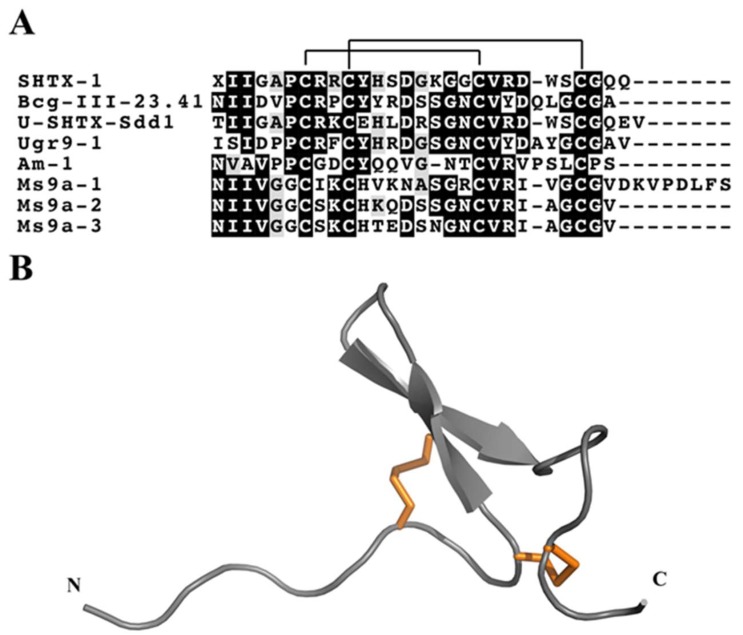
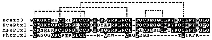
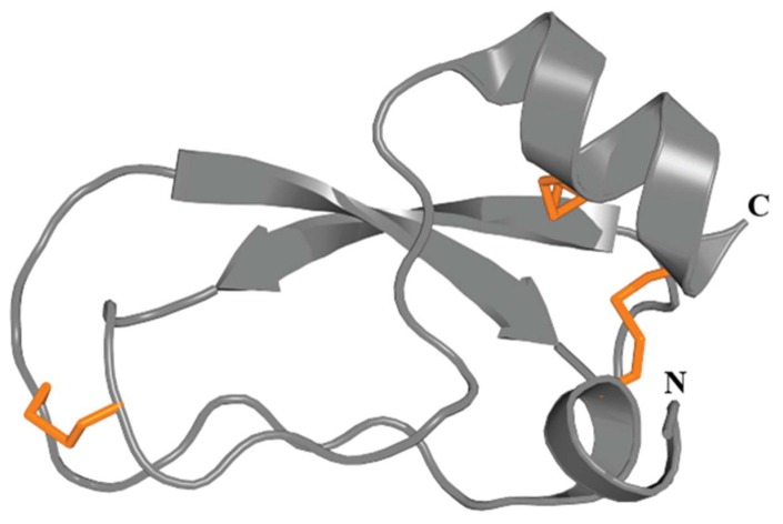
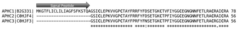
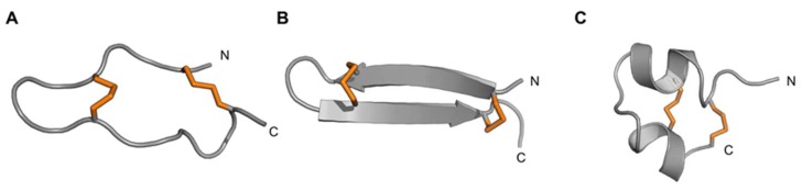
Sea anemones produce venoms of exceptional molecular diversity, with at least 17 different molecular scaffolds reported to date. These venom components have traditionally been classified according to pharmacological activity and amino acid sequence. However, this classification system suffers from vulnerabilities due to functional convergence and functional promiscuity. Furthermore, for most known sea anemone toxins, the exact receptors they target are either unknown, or at best incomplete. In this review, we first provide an overview of the sea anemone venom system and then focus on the venom components. We have organised the venom components by distinguishing firstly between proteins and non-proteinaceous compounds, secondly between enzymes and other proteins without enzymatic activity, then according to the structural scaffold, and finally according to molecular target.

Keywords: cytotoxin; enzyme; molecular scaffold; neurotoxin; sea anemone; toxin; venom.


Conflict of interest statement

The authors declare no conflict of interest

Figures

Figure 1
Figure 1
(A) Phylogenetic tree of Cnidarians. Representative Medusozoa depicted here are: Hydra viridis (Hydrozoa), Aurelia aurita (Scyphozoa), Chironex fleckeri (Cubozoa), and Haliclystus sp (Staurozoa). Representative Anthozoa are the sea anemone Nematostella vectensis (left) and the coral Acropora millepora (right). Figure modified with permission from Marine Genomics; published by Elsevier, 2015 [9]. (B) Phylogenetic relationships among major lineages of sea anemones (after Rodrigues et al., [4]).
Figure 2
Figure 2
3D structures of actinoporins as exemplified by (A) Δ-stichotoxin-She1a (Sticholysin I; PDB accession code 2KS4) and (B) Δ-actitoxin-Aeq1a (Equinatoxin II; PDB 1KD6). The N- and C-termini are labelled.
Figure 3
Figure 3
Representative sea anemone toxins with an ATX III motif. (A) Alignment of representative sea anemone toxins that adopt the ATX III motif. Disulfide bridge connectivities are indicated above the sequence alignment. Amino acid identities (black boxes) and similarities (grey boxes) are shown. The representative toxin sequences shown are ATX III (δ-AITX-Avd2a; UniProt P01535), Av7 (δ-AITX-Avd2b 3; UniProt C3TS10), and Av10 (δ-AITX-Avd2c; UniProt C3TS07). (B) 3D structure of the sea anemone toxin Av3 (PDB accession code 1ANS). The three disulfide bonds are represented by orange tubes.
Figure 4
Figure 4
Sequence and structure of sea anemone toxins that adopt a β-defensin fold. (A) Representative 3D structures of sea anemone toxins that adopt a β-defensin fold. Disulfide bonds are shown as orange tubes. Toxins are grouped according to their molecular targets. (B) Alignment of representative sea anemone toxins that contain a β-defensin motif. Disulfide connectivities are indicated above the sequence alignment. Identical and similar amino acid residues are highlighted in black and grey, respectively. The representative toxin sequences shown are AP-B (Δ-actitoxin-Axm1b, UniProt P01531), ShI (Δ-stichotoxin-She1a, UniProt P19651), CgNa (Δ-actitoxin-Cgg1a, UniProt P0C280), BDS1 (Δκ-actitoxin-Avd4a, UniProt P11494), APETx1 (κ-actitoxin-Ael2a, UniProt P61541), APETx2 (π-actitoxin-Ael2b, UniProt P61542).
Figure 5
Figure 5
Superimposed cartoon representation of the structures of CgNa (Δ-actitoxin-Cgg1a) and ApB (left) and CgNa and ShI (Δ-stichotoxin-She1a) (right). CgNa is coloured green and dark grey. Figure modified with permission from Biochemical Journal; published by Portland Press, 2007 [118].
Figure 6
Figure 6
Representative sea anemone toxins with BBH mortif. (A) Alignment of representative sea anemone toxins that adopt the BBH motif. Disulfide connectivities are indicated above the sequence alignment. Amino acid identities (black boxes) and similarities (grey boxes) are highlighted. The representative toxin sequences shown are SHTX-1 (κ-stichotoxin-Shd1a; UniProt P0C7W7), Bcg-III-23.41 (U-BcgTx1a; UniProt P86467), U-SHTX-Sdd1 (UniProt C0HJB4), Ugr9-1 (π-AnmTX Ugr 9a-1; UniProt R4ZCU1), Am-1 (Δ-AITX-Amc1a; UniProt P69929), Ms9a-1 (T-AnmTX Ms 9a-1; UniProt C0HK13), Ms9a-2 (T-AnmTX Ms 9a-2; UniProt C0HK13) and Ms9a-3 (T-AnmTX Ms 9a-3; UniProt C0HK13). (B) 3D structure of the sea anemone toxin Ugr9-1 (PDB 2LZO). The two disulfide bonds are represented by orange tubes.
Figure 7
Figure 7
Alignment of sea anemone toxins that likely adopt an ICK fold. Predicted disulfide connectivities are indicated. Amino acid identities (black boxes) and similarities (grey boxes) are highlighted. The representative toxin sequences shown are BcsTx3 (κ-actitoxin-Bcs4a; UniProt C0HJC4), NvePTx1 (U-EWTX-NvePTx1; UniProt A7RMN1), MsePTx1 (U-metritoxin-Msn2a; UniProt P0DMD7) and PhcrTx1 (π-phymatoxin-Pcf1a; UniProt C0HJB1).
Figure 8
Figure 8
3D structure of the KV type 2 sea anemone toxin ShPI-I (π-stichotoxin-She2a) (PDB 3OFW). Peptides of this type are homologous to Kunitz-type serine protease inhibitors. The three disulfide bonds are represented by orange tubes.
Figure 9
Figure 9
Aligment of APHC1, 2 and 3 (UniProt B2G331, C0HJF3 and C0HJF4, respectively). Cysteines are highlighted in bold, sequence differences are marked with dots, and conserved positions are marked with *.
Figure 10
Figure 10
Comparison of the PHAB fold with other peptide folds containing two disulfide bonds and a similar number of residues (16–29 residues). Disulfide bonds are shown as orange tubes and N- and C-termini are labelled. (A) Ate1a (PDB 6AZA); (B) β-hairpin fold represented by the spider peptide gomesin (PDB 1KFP); (C) CS α/α motif represented by the scorpion toxin κ-hefutoxin1 (PDB 1HP9). Figure modified with permission from Cellular and Molecular Life Sciences; published by Springer International Publishing, 2018 [148].
Figure 11
Figure 11
Representative sea anemone toxins with SCRiP motif. (A) Alignment of representative sea anemone SCRiP toxins. Disulfide connectivities are indicated above the sequence alignment. Amino acid identities (black boxes) and similarities (grey boxes) are highlighted. The representative toxin sequences shown are Ueq12-1 (τ-AnmTx Ueq 12-1; UniProt C0HK26), AvirSCRiP (UniProt P0DL61) and MsenSCRiP (UniProt P0DL60). (B) 3D structure of the sea anemone toxin Ueq 12-1 (PDB 5LAH). The three disulfide bonds are shown as orange tubes.
Figure 12
Figure 12
3D structure of KV type 1 sea anemone toxins. (A) Structures of BgK (κ-actitoxin-Bgr1a, PDB 1BGK), and (B) ShK (PDB 1ROO). The three disulfide bonds are represented by orange tubes.

References

    1. Brusca R.C., Brusca G.J., Haver N. Invertebrates. 2nd ed. Sinauer Associates; Sunderland, MA, USA: 2003.
    1. Stewart Z.K., Pavasovic A., Hock D.H., Prentis P.J. Transcriptomic investigation of wound healing and regeneration in the cnidarian Calliactis polypus. Sci. Rep. 2017;7:41458. doi: 10.1038/srep41458. - DOI - PMC - PubMed
    1. Galliot B., Schmid V. Cnidarians as a model system for understanding evolution and regeneration. Int J. Dev. Biol. 2002;46:39–48. - PubMed
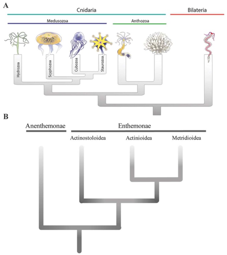
    1. Rodriguez E., Barbeitos M.S., Brugler M.R., Crowley L.M., Grajales A., Gusmao L., Haussermann V., Reft A., Daly M. Hidden among sea anemones: the first comprehensive phylogenetic reconstruction of the order Actiniaria (Cnidaria, Anthozoa, Hexacorallia) reveals a novel group of hexacorals. PLoS One. 2014;9:e96998. doi: 10.1371/journal.pone.0096998. - DOI - PMC - PubMed
    1. Shick J.M. A Functional Biology of Sea Anemones. Chapman & Hall; London, UK: 1991.

LinkOut - more resources