Review
. 2019 Jun 3;146(11):dev150920.
doi: 10.1242/dev.150920.

The endoderm: a divergent cell lineage with many commonalities

Affiliations
Review

The endoderm: a divergent cell lineage with many commonalities

Sonja Nowotschin et al. Development. .

Abstract

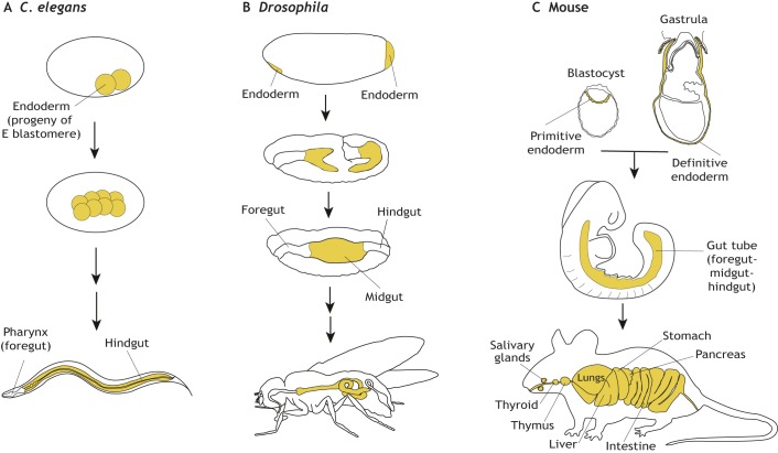
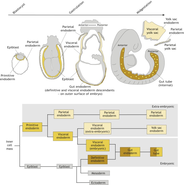
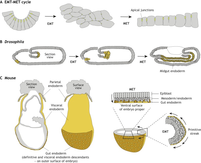
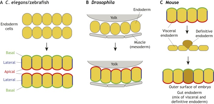
The endoderm is a progenitor tissue that, in humans, gives rise to the majority of internal organs. Over the past few decades, genetic studies have identified many of the upstream signals specifying endoderm identity in different model systems, revealing them to be divergent from invertebrates to vertebrates. However, more recent studies of the cell behaviours driving endodermal morphogenesis have revealed a surprising number of shared features, including cells undergoing epithelial-to-mesenchymal transitions (EMTs), collective cell migration, and mesenchymal-to-epithelial transitions (METs). In this Review, we highlight how cross-organismal studies of endoderm morphogenesis provide a useful perspective that can move our understanding of this fascinating tissue forward.

Keywords: Collective cell migration; Endoderm; Epithelial-to-mesenchymal transitions; Mesenchymal-to-epithelial transitions; Morphogenesis.


Conflict of interest statement

Competing interestsThe authors declare no competing or financial interests.

Figures

Fig. 1.
Fig. 1.
Location of the endoderm throughout the development of worm, Drosophila and mouse embryos. (A) In C. elegans, the endoderm is derived from the progeny of the E blastomere and gives rise to endodermal cells of the entire gut tube. (B) In Drosophila, endoderm forms at the anterior and posterior poles of the embryo and then invaginates to form the midgut. The foregut and hindgut of the gut tube are of ectodermal origin. (C) In mice, the entire gut tube is composed of cells of two different endodermal origins – (1) the extra-embryonic endoderm, which comprises visceral endoderm descendants of the primitive endoderm specified in the pre-implantation blastocyst; and (2) embryonic endoderm (usually referred to as definitive endoderm) – which are descendants of the epiblast, specified at gastrulation. The gut tube then gives rise to the epithelial lining of all endodermal organs of the adult mouse.
Fig. 2.
Fig. 2.
The endoderm of the mouse embryo arises from two sources with distinct developmental origins. Schematic overview (top) and lineage tree (bottom) depicting the development of endodermal tissues in mice, from the blastocyst stage to the midgestation embryo. The gut endoderm forms on the surface of the embryo at gastrulation where definitive endoderm (derived from the epiblast; brown) cells intercalate (egress; Schöck and Perrimon, 2002) into the overlying visceral endoderm (derived from the primitive endoderm, yellow). The gut endoderm then becomes internalized and forms the gut tube and will give to the epithelial lining of all endodermal tissues in the adult organism. The gut tube therefore comprises cells of two different origins: extra-embryonic (beige) and embryonic (yellow). Parietal and yolk sac endoderm (beige), which are also derived from primitive endoderm in the blastocyst, solely give rise to extra-embryonic structures.
Fig. 3.
Fig. 3.
The EMT-migration/translocation/relocation-MET cycle in Drosophila and mouse embryos. (A) Schematic of cells undergoing an EMT-MET cycle. Cells within an epithelium undergoing EMT lose their apico-basal polarity and loosen their cell-cell junctions (green). After undergoing EMT, cells display a mesenchymal morphology and adopt migratory behaviour. As they rejoin an epithelium and undergo MET, they re-establish cell polarity (by upregulating apico-basal polarized proteins) and reform or reinforce cell-cell junctions. (B) In Drosophila, an EMT-MET cycle occurs during midgut formation. Future endoderm cells delaminate from the anterior and posterior poles of the embryo. Cells from both sections migrate and meet to form the midgut section of the gut. Through interactions with the underlying mesoderm, endoderm cells undergo MET and repolarize to form the midgut epithelium. (C) In mice, an EMT-MET cycle occurs during gastrulation. Definitive endoderm cells (brown) undergo EMT when they leave the primitive streak and migrate along the wings of mesoderm. They then undergo MET as they intercalate in the overlying visceral endoderm (yellow) epithelium to form the gut endoderm. Section and surface whole-mount views show the location of the gut endoderm on the surface of the mouse embryo, consisting of definitive endoderm cells and visceral endoderm cells. Cross-sections through the embryo show the position of an EMT at the primitive streak, and an MET during gut endoderm formation.
Fig. 4.
Fig. 4.
Endoderm repolarization: the different ways cells can re-polarize. (A) In C. elegans and zebrafish, the endoderm comprises a rod of cells that localizes apico-basal polarity proteins at its centre and subsequently generates multiple small lumens, which coalesce into a single one. (B) In Drosophila, the endoderm comprises a cup-shaped sheet of cells acquiring apico-basal polarity through cell-cell and cell-basement membrane interactions. (C) In mice, definitive endoderm cells (brown) repolarize as they intercalate into the overlying visceral endoderm (yellow) epithelium. During the intercalation process, visceral endoderm cells relax their apico-basal polarity and cell-cell junctions to facilitate definitive endoderm intercalation. However, once definitive endoderm cells have egressed into the visceral endoderm epithelium, both cell types coordinately repolarize and re-establish cell-cell junctions.

References

    1. Achilleos A., Wehman A. M. and Nance J. (2010). PAR-3 mediates the initial clustering and apical localization of junction and polarity proteins during C. elegans intestinal epithelial cell polarization. Development 137, 1833-1842. 10.1242/dev.047647 - DOI - PMC - PubMed
    1. Alev C., Wu Y., Kasukawa T., Jakt L. M., Ueda H. R. and Sheng G. (2010). Transcriptomic landscape of the primitive streak. Development 137, 2863-2874. 10.1242/dev.053462 - DOI - PubMed
    1. Alvers A. L., Ryan S., Scherz P. J., Huisken J. and Bagnat M. (2014). Single continuous lumen formation in the zebrafish gut is mediated by smoothened-dependent tissue remodeling. Development 141, 1110-1119. 10.1242/dev.100313 - DOI - PMC - PubMed
    1. Andrew D. J. and Ewald A. J. (2010). Morphogenesis of epithelial tubes: Insights into tube formation, elongation, and elaboration. Dev. Biol. 341, 34-55. 10.1016/j.ydbio.2009.09.024 - DOI - PMC - PubMed
    1. Arnold S. J. and Robertson E. J. (2009). Making a commitment: cell lineage allocation and axis patterning in the early mouse embryo. Nat. Rev. Mol. Cell Biol. 10, 91-103. 10.1038/nrm2618 - DOI - PubMed

Publication types