. 2019 Jun 18;116(25):12167-12172.
doi: 10.1073/pnas.1900172116. Epub 2019 Jun 3.

Ratiometric two-photon microscopy reveals attomolar copper buffering in normal and Menkes mutant cells

Affiliations

Ratiometric two-photon microscopy reveals attomolar copper buffering in normal and Menkes mutant cells

M Thomas Morgan et al. Proc Natl Acad Sci U S A. .

Abstract

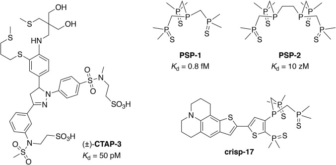
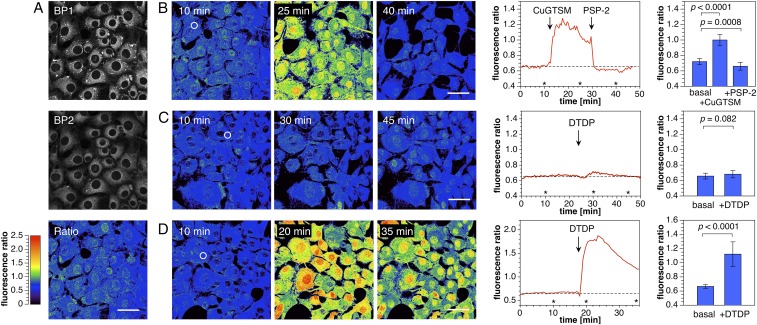
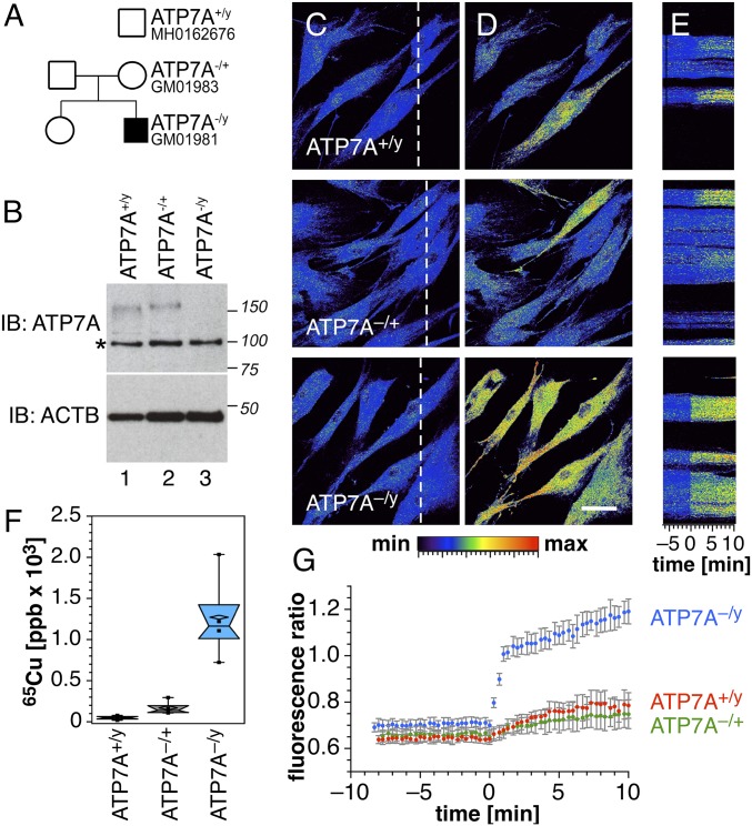
Copper is controlled by a sophisticated network of transport and storage proteins within mammalian cells, yet its uptake and efflux occur with rapid kinetics. Present as Cu(I) within the reducing intracellular environment, the nature of this labile copper pool remains elusive. While glutathione is involved in copper homeostasis and has been assumed to buffer intracellular copper, we demonstrate with a ratiometric fluorescent indicator, crisp-17, that cytosolic Cu(I) levels are buffered to the vicinity of 1 aM, where negligible complexation by glutathione is expected. Enabled by our phosphine sulfide-stabilized phosphine (PSP) ligand design strategy, crisp-17 offers a Cu(I) dissociation constant of 8 aM, thus exceeding the binding affinities of previous synthetic Cu(I) probes by four to six orders of magnitude. Two-photon excitation microscopy with crisp-17 revealed rapid, reversible increases in intracellular Cu(I) availability upon addition of the ionophoric complex CuGTSM or the thiol-selective oxidant 2,2'-dithiodipyridine (DTDP). While the latter effect was dramatically enhanced in 3T3 cells grown in the presence of supplemental copper and in cultured Menkes mutant fibroblasts exhibiting impaired copper efflux, basal Cu(I) availability in these cells showed little difference from controls, despite large increases in total copper content. Intracellular copper is thus tightly buffered by endogenous thiol ligands with significantly higher affinity than glutathione. The dual utility of crisp-17 to detect normal intracellular buffered Cu(I) levels as well as to probe the depth of the labile copper pool in conjunction with DTDP provides a promising strategy to characterize perturbations of cellular copper homeostasis.

Keywords: Menkes disease; copper homeostasis; fluorescent probes; glutathione; two-photon microscopy.


Conflict of interest statement

The authors declare no conflict of interest.

Figures

Fig. 1.
Fig. 1.
Structure of the lipid-compatible fluorescent indicator CTAP-3 and Cu(I)-selective reagents utilizing phosphine sulfide-stabilized phosphine (PSP) coordination motifs, including ligands PSP-1 and PSP-2, and the Cu(I)-indicator crisp-17.
Fig. 2.
Fig. 2.
Synthesis of the Cu(I)-responsive indicator crisp-17 and Cu(I)-insensitive control fluorophore crisp-17ctrl.
Fig. 3.
Fig. 3.
Spectral response and selectivity of crisp-17 toward Cu(I) (supplied as [Cu(I)MCL-2]PF6). (A) UV-vis absorption spectra of crisp-17 (5 µM) in 95% MeOH-5% H2O during titration with Cu(I) (0–6.5 µM). (B) Fluorescence emission spectra (λex = 460 nm) under the same conditions. (Inset) Emission intensity at 560 nm vs. molar ratio of Cu(I) to probe. (C) Emission spectra (λex = 450 nm) of crisp-17 (2 µM) equilibrated with liposome suspension (4:1 POPC–POPG, 100 µM total lipids, 10 mM Pipes, pH 7.0, 0.1 M KCl, 25 °C) in the absence (blue trace) and presence (red trace) of 4 µM Cu(I). (D) Ratio of the integrated emission intensities F(590–750 nm)/F(480–580 nm) of crisp-17 (2 µM) in the presence of biologically relevant transition metal ions (10 µM) before (blue bars) and after (green bars) addition of 4 µM Cu(I) (liposome and buffer composition as in C). Probe response in the absence of a competing metal ion.
Fig. 4.
Fig. 4.
Fluorimetric Cu(I) competition titrations of crisp-17 against MCL-1 and GSH (4:1 DMPC–DMPG, 100 µM total lipids, 10 mM Pipes, pH 7.0, 0.1 M KCl, 25 °C, λex = 450 nm). (A) crisp-17 (1 µM) was equilibrated with 10 µM [Cu(I)MCL-1]PF6 (red trace) at pH 7.0 and subsequently titrated with MCL-1 (10–640 µM, black traces). Full reversibility was confirmed by adding 10 µM PSP-2 (blue trace). Nonlinear least-squares fitting yielded logK of 17.0 ± 0.1. (B) Fit quality evaluated at 530 nm. (C) Apparent fractional saturation of crisp-17, calculated as (IIfree)/(IboundIfree), where I represents integrated fluorescence intensity from 500 to 550 nm, upon titration with Cu(I) (as [Cu(I)MCL-2]PF6) in the presence of 4 mM GSH at pH 7.0. Least-squares fitting (red line) yielded logK = 17.1 ± 0.1. (D) Calculated fraction of crisp-17 bound to Cu(I) and of total Cu(I) bound to GSH versus thermodynamically available Cu(I) (pCu = −log[Cu+aq]). The dashed line and blue shading demarcate the Cu(I) availability corresponding to 10% fractional saturation of the probe. The red-shaded area indicates the GSH Cu(I) buffer window within the cytosolic pH range.
Fig. 5.
Fig. 5.
Two-photon ratiometric imaging of labile copper dynamics in live mouse fibroblasts stained with crisp-17 (1 µM). (A, Top) Fluorescence intensity images acquired with emission channels of 479–536 nm (BP1) and 611–750 nm (BP2) with two-photon excitation at 880 nm. (A, Bottom) Ratio image with ratio = BP2/BP1. (B, Left) Ratio images before and after addition of 10 µM CuGTSM. (Scale bars, 50 μm.) (B, Middle) Time course of the average ratio change for the region of interest (ROI) indicated with a white circle in the Left. The asterisks indicate time points for the respective ratio images shown to the Left. (B, Right) Mean fluorescence ratio of the cytoplasmic region averaged over 20 cells (P values calculated for n = 20 using a two-tailed test). (C) Experiment as described for B but with addition of 1 mM 2,2′-dithiodipyridine (DTDP) in lieu of CuGTSM. (D) Experiment as described for C but employing cells grown in medium supplemented with 50 µM CuCl2.
Fig. 6.
Fig. 6.
Copper phenotypes of Menkes disease fibroblasts. (A) Pedigrees of a family affected by Menkes disease. (B) ATP7A-null subject GM01981 was confirmed by immunoblot analysis. Asterisk denotes nonspecific band recognized by the antibody. Loading controls were performed with anti–β-actin antibodies (ACTB). (C) Wild-type (ATP7A+/y), carrier mother (ATP7A−/+, GM01983), and Menkes son (ATP7A−/y, GM01981) fibroblasts were imaged by two-photon microscopy with crisp-17. Color scale depicts copper-dependent emission ratio (0–2.5 range) at 570–640 nm/500–550 nm. (D) Copper was mobilized by the addition of the oxidizing reagent DTDP (100 µM). (Scale bar, 50 µm.) (E) Kymographs of cells in C (dashed line) with time 0 corresponding to the addition of DTDP. (F) Copper content phenotype confirmed by inductively coupled plasma mass spectrometry (one-way ANOVA followed by Dunnett's multiple comparisons; n = 3). (G) Fluorescence emission ratios before and after DTDP addition. Average ± SE from six independent experiments.

References

    1. Öhrvik H., Aaseth J., Horn N., Orchestration of dynamic copper navigation—new and missing pieces. Metallomics 9, 1204–1229 (2017). - PubMed
    1. Lutsenko S., Human copper homeostasis: A network of interconnected pathways. Curr. Opin. Chem. Biol. 14, 211–217 (2010). - PMC - PubMed
    1. Robinson N. J., Winge D. R., Copper metallochaperones. Annu. Rev. Biochem. 79, 537–562 (2010). - PMC - PubMed
    1. Herd S. M., Camakaris J., Christofferson R., Wookey P., Danks D. M., Uptake and efflux of copper-64 in Menkes’-disease and normal continuous lymphoid cell lines. Biochem. J. 247, 341–347 (1987). - PMC - PubMed
    1. Morgan M. T., Bagchi P., Fahrni C. J., “Fluorescent probes for monovalent copper” in Metals in Cells, Culotta V, Scott RA, Eds. (Wiley, 2013), pp. 65–84.

Publication types

LinkOut - more resources