. 2019 Jun 3;10(1):2385.
doi: 10.1038/s41467-019-10363-1.

Structures of BCL-2 in complex with venetoclax reveal the molecular basis of resistance mutations

Affiliations

Structures of BCL-2 in complex with venetoclax reveal the molecular basis of resistance mutations

Richard W Birkinshaw et al. Nat Commun. .

Abstract

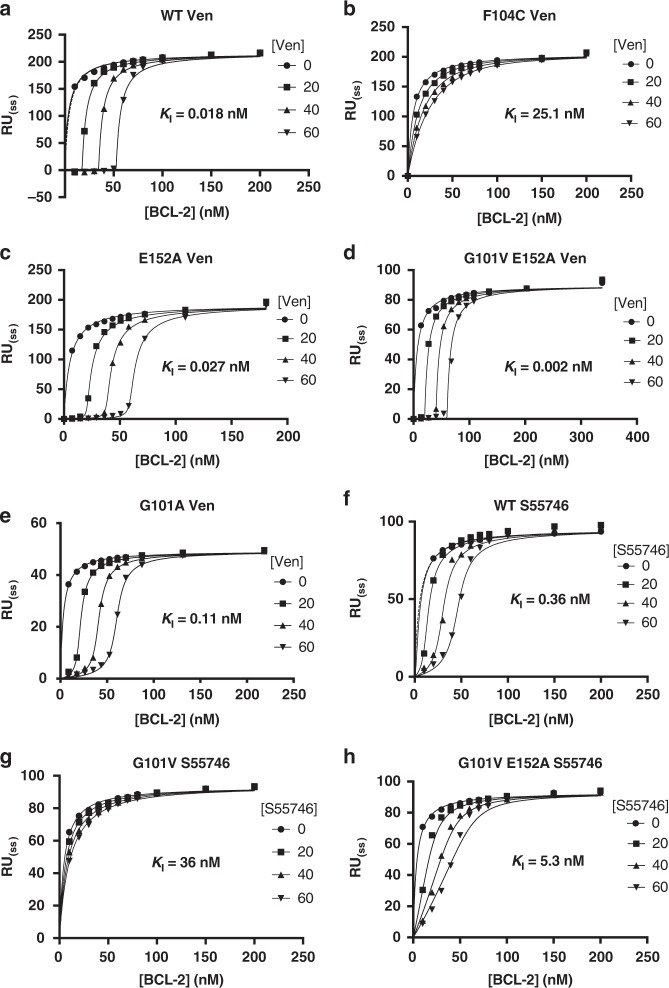
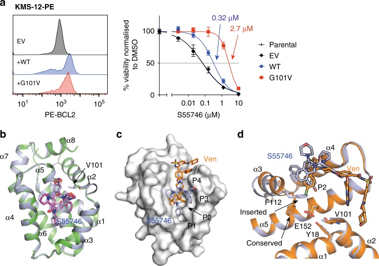
Venetoclax is a first-in-class cancer therapy that interacts with the cellular apoptotic machinery promoting apoptosis. Treatment of patients suffering chronic lymphocytic leukaemia with this BCL-2 antagonist has revealed emergence of a drug-selected BCL-2 mutation (G101V) in some patients failing therapy. To understand the molecular basis of this acquired resistance we describe the crystal structures of venetoclax bound to both BCL-2 and the G101V mutant. The pose of venetoclax in its binding site on BCL-2 reveals small but unexpected differences as compared to published structures of complexes with venetoclax analogues. The G101V mutant complex structure and mutant binding assays reveal that resistance is acquired by a knock-on effect of V101 on an adjacent residue, E152, with venetoclax binding restored by a E152A mutation. This provides a framework for considering analogues of venetoclax that might be effective in combating this mutation.


Conflict of interest statement

R.W.B., J.-N.G., C.S.L., D.L., C.A.W., M.A.A., G.L., I.J.M., R.T., A.W.R., D.C.S.H., P.M.C. and P.E.C. are employees of the Walter and Eliza Hall Institute, which has an agreement with Genentech and AbbVie and receives milestone and royalty payments related to venetoclax. Employees of Walter and Eliza Hall Institute may be eligible for financial benefits related to these payments. J.-N.G., M.A.A., G.L., I.J.M., A.W.R., D.C.S.H., P.M.C. and P.E.C. receive such a financial benefit as a result of previous research related to venetoclax. G.L., D.C.S.H., P.M.C. and P.E.C. have received research funding from Genentech. A.W.R has received research funding from AbbVie. The remaining author declares no competing interests.

Figures

Fig. 1
Fig. 1
Venetoclax binding to BCL-2. a Chemical structures of venetoclax (Ven) and related compounds ABT-263 and compound 1. Key features are marked, including the benzamide (BA), the 4-chlorophenyl (CP), the partially saturated 4–4-dimethylcyclohex-1-ene (4DM), the 5–5-dimethylcyclohex-1-ene (5DM), the saturated 4′-chlorobiphenyl (BP), the azaindole (AI) and phenyl sulfonyl (PS). b Crystal structures showing binding of the two venetoclax conformers (Ven A and Ven B, orange), and comparison to ABT-263 (green, PDB id 4LVT) and compound 1 (cyan, PDB id 4MAN) binding to the BCL-2 groove. BCL-2 is shown with surface representation (white) and P2 and P4 pockets indicated along with arrows depicting changes between venetoclax alternate conformations for BA and 4DM moieties. c, d overlays of venetoclax with ABT-263 (c) and compound 1 (d) with arrows indicating deviations compared to the venetoclax 4DM above the P2 pocket
Fig. 2
Fig. 2
BCL-2 mutations and venetoclax binding. a location of G101V and F104L mutants relative to the BCL-2 groove, α1–8 helices and P2 (orange) and P4 (blue) pockets. Pockets volumes were defined by venetoclax (Ven) chlorophenyl (CP) for P2 pocket and azaindole ring for P4 pocket, see methods for further details. b, c Overlay of BCL-2 WT and BCL-2 G101V bound to venetoclax. d Overlay of BCL-2 WT and BCL-2 F104L bound to venetoclax. e Surface representation of the BCL-2 WT:venetoclax P2 pocket. f Surface representation of the BCL-2 F104L:venetoclax P2 pocket. In a–f Venetoclax is shown in orange for BCL-2 WT and yellow in mutants, BCL-2 WT in green, BCL-2 G101V in pale blue, F104L in purple and surfaces in white. Arrows indicate key residues, dashed arrows indicate residue movements or movement of venetoclax
Fig. 3
Fig. 3
Steady-state competition SPR with BCL-2 mutants, BIMBH3 and venetoclax or S55746. SPR chip surfaces were immobilised with BIMBH3 peptide and analytes with the indicated BCL-2 mutants pre-combined with either ae venetoclax (Ven) or fh S55746 at the indicated concentrations (circles 0 nM, squares 20 nM, triangles point up 40 nM, triangles point down 60 nM). Response at steady-state is plotted against BCL-2 concentration, with fits from a steady-state competition SPR model shown (see methods and Supplementary Figs. 5, 6 for further details), that were used to derive the indicated mean KI values for venetoclax or S55746 binding to BCL-2. Data are representative of at least n = 2 independent experiments
Fig. 4
Fig. 4
BCL-2 G101V binding to S55746. a Expression of BCL-2 G101V mutant reduces S55746 sensitivity in the KMS-12-PE B-lineage cell line. WT and G101V mutant BCL-2 proteins were expressed at similar levels in KMS-12-PE cells as demonstrated by the FACS profiles. In vitro sensitivity to S55746 (0–10 μM) was measured after 24 h by a CellTiter-Glo assay. Data are representative of n = 3 independent experiments with mean LC50 values ± SD (1 Standard Deviation) for empty vehicle (EV, black), WT (blue) and G101V (red) indicated. b Overlay of the published BCL-2:S55746 structure (green and magenta, PDB id 6GL8) with the BCL-2 G101V:S55746 structure (light blue) showing consistency in S55746 orientation. c, d Overlay of BCL-2 G101V structures bound to S55746 (light blue) and venetoclax (Ven, orange). c BCL-2 protein surface displayed from the BCL-2 G101V:S55746 structure showing P1–4 pockets. d Cartoon representation with key residues indicated in stick representation, inserted conformation of F112 in the BCL-2 G101V—S55746 and WT conformation from BCL-2 G101V:venetoclax structure are indicated
Fig. 5
Fig. 5
BCL-2 surface contacts with either venetoclax, S55746 or BaxBH3 peptide. Structure of BCL-2 G101V with a Venetoclax (Ven) or b S55746, or c BCL-2 WT with a BaxBH3 peptide. BCL-2 surface contacts are coloured according to interactions with venetoclax (orange), S55746 (magenta) or BaxBH3 peptide (slate). Key BCL-2 binding pockets (P1 through P4) indicated for each compound and hydrophobic (ϕ) 1–4 residues (L, L, I, L) from the BaxBH3 peptide. c BCL-2 surface contacts with venetoclax are coloured in orange and with BaxBH3 peptide in slate highlighting the increased surface contact area of BaxBH3 relative to venetoclax (PDB id 2XA0)

References

    1. Vaux DL, Cory S, Adams JM. Bcl-2 gene promotes haemopoietic cell survival and cooperates with c-myc to immortalize pre-B cells. Nature. 1988;335:440–442. doi: 10.1038/335440a0. - DOI - PubMed
    1. Czabotar PE, Lessene G, Strasser A, Adams JM. Control of apoptosis by the BCL-2 protein family: implications for physiology and therapy. Nat. Rev. Mol. Cell Biol. 2014;15:49–63. doi: 10.1038/nrm3722. - DOI - PubMed
    1. Cory S, Roberts AW, Colman PM, Adams JM. Targeting BCL-2-like proteins to kill cancer cells. Trends Cancer. 2016;2:443–460. doi: 10.1016/j.trecan.2016.07.001. - DOI - PubMed
    1. Roberts AW, et al. Targeting BCL2 with venetoclax in relapsed chronic lymphocytic leukemia. N. Engl. J. Med. 2016;374:311–322. doi: 10.1056/NEJMoa1513257. - DOI - PMC - PubMed
    1. Sattler M, et al. Structure of Bcl-xL-Bak peptide complex: recognition between regulators of apoptosis. Science. 1997;275:983–986. doi: 10.1126/science.275.5302.983. - DOI - PubMed

Publication types

MeSH terms