. 2019 Sep;4(9):1571-1581.
doi: 10.1038/s41564-019-0466-x. Epub 2019 Jun 3.

Complex N-glycan breakdown by gut Bacteroides involves an extensive enzymatic apparatus encoded by multiple co-regulated genetic loci

Affiliations

Complex N-glycan breakdown by gut Bacteroides involves an extensive enzymatic apparatus encoded by multiple co-regulated genetic loci

Justina Briliūtė et al. Nat Microbiol. 2019 Sep.

Abstract

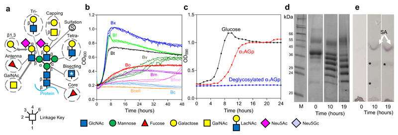
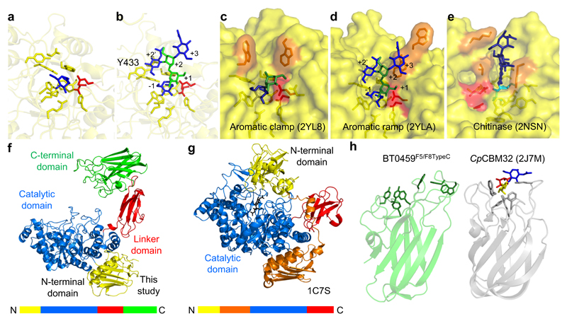
Glycans are the major carbon sources available to the human colonic microbiota. Numerous N-glycosylated proteins are found in the human gut, from both dietary and host sources, including immunoglobulins such as IgA that are secreted into the intestine at high levels. Here, we show that many mutualistic gut Bacteroides spp. have the capacity to utilize complex N-glycans (CNGs) as nutrients, including those from immunoglobulins. Detailed mechanistic studies using transcriptomic, biochemical, structural and genetic techniques reveal the pathway employed by Bacteroides thetaiotaomicron (Bt) for CNG degradation. The breakdown process involves an extensive enzymatic apparatus encoded by multiple non-adjacent loci and comprises 19 different carbohydrate-active enzymes from different families, including a CNG-specific endo-glycosidase activity. Furthermore, CNG degradation involves the activity of carbohydrate-active enzymes that have previously been implicated in the degradation of other classes of glycan. This complex and diverse apparatus provides Bt with the capacity to access the myriad different structural variants of CNGs likely to be found in the intestinal niche.


Conflict of interest statement

Competing interests

The authors declare no competing interests

Figures

Figure 1
Figure 1. Complex N-glycans as a nutrient source in Bacteroides species
a, Structure of a biantennary complex N-glycan (CNG) with other possible decorations and variations. The linkage is indicated by the key, however the sialic acid sugars can be attached through α2,3 or 2,6 linkages to the galactose and also to the antennary GlcNAc. Biantennary CNG have both antennae linked through β1,2 bonds and additional antenna have either a β1,4-linkage (on the α1,3 arm) or a β1,6-linkage (on the α1,6 arm) to produce tri- and tetra-antennary CNG, respectively. Common modifications to this core model are shown in dotted circles. b, Growth on native α1AGp of different Bacteroides spp. - B. thetaiotaomicron (Bt, black), B. xylanisolvans (Bx, dark blue), B. ovatus (Bo, red), B. vulgatus (Bv, dark green), B. finegoldii (Bfine, pink), B. massiliensis (Bm, magneta), B. fragilis (Bf, light green), B. caccae (Bc, light blue) and B. cellulolyticus (Bcell, orange). Growth curves were carried out at least twice for each species. c, Growth of B. thetaiotaomicron on glucose (5 mg/ml), α1AGp and deglycosylated α1AGp (both 20mg/ml) as the sole carbon source. d, Supernatant samples were taken at the start, 10 hour and 19 hour time points from the growth of Bt on α1AGp show in panel c and were analysed by SDS-PAGE. Fully glycosylated α1AGp is shown at T0 and fully deglycosylated α1AGp is the bottom 23.5 kDa protein band in the 10 h and 19 h time point lanes. e, TLC of the cell free spent media at the 10 h and 19 h time points to analyse glycans present. The top band is sialic acid (white arrow and “SA”) and two other faint glycan bands are indicated to (*). The results are representative of at least three independent replicates. Full versions of the gels and TLCs can be found in Supplementary Figure 1 and 23.
Figure 2
Figure 2. Genes upregulated in Bt genes during growth on CNG
a, A schematic of the 6 CAZyme-containing loci upregulated in Bt during growth on α1AGp. TM: transmembrane protein; pSGBP: putative surface glycan binding protein. b, A model of the degradation of CNGs by Bt. BT1625GH29 is placed in the periplasm but could in fact be facing the outside of the cell. The degradation of the common core tetrasaccharide likely occurs through the activity of previously characterised enzymes located in the periplasm and cytoplasm (Supplementary Table 2, Supplemental Results and Discussion).
Figure 3
Figure 3. The degradation of biantennary CNG by recombinant enzymes from Bt
a, α1AGp digested with BT1044GH18 endo-GlcNAc’ase only and b, then in combination with BT0455GH33 sialidase, and c, then also BT0461GH2 β-galactosidase. d-f, Each of the assays shown in a-c were stopped after a 24 h incubation using heat denaturation and BT1035GH163 then added. The time shown for the different chromatograms varies between panels to provide clarity of the main peaks. The glycan products were labelled with procainamide and analysed by LC-FLD-ESI-MS (see Materials and Methods). The most abundant glycan products are annotated on the chromatograms and the minor products are shown in the key at the bottom. The same glycan species detected in multiple peaks is likely due to different linkages and glycosylation on different arms, which cannot be determined by the analytical methods employed. Neu5Ac and Neu5Gc are pink and light blue, respectively. The linkages they are attached through could not be determined using the techniques employed here and also likely vary between glycans and glycoproteins. The results are representative of at least three independent replicates. A sugar key is included in Figure 1.
Figure 4
Figure 4. The activity of BT1044GH18 on immunoglobulin substrates and structural of the enzyme
Activity of BT1044GH18 against a, human serum IgG b, human serum IgA and c, human colostrum IgA is shown in the presence of BT0455GH33 sialidase. The products with a β1,4 bisecting GlcNAc are indicated to by a white asterisk. The results are representative of at least three independent replicates. d, Cartoon depiction of the predicted orientation of BT1044GH18 on the cell surface of Bt showing the attachment to the cell surface through a lipid anchor. Loops 1-7 on the surface of the enzyme with the active site are coloured yellow, green, orange, blue, magenta, purple and dark green, respectively. e, The surface of the BT1044GH18 crystal structure is shown again with the same loop colouring and the two catalytic residues are in red. For comparison, the previously published crystal structure of the CNG-active enzyme EndoF3 (1EOM) from Elizabethkingia meningoseptica is shown (cyan). The conserved residues from the DxxDxDxE motif have been coloured orange and the approximate +1 (GlcNAc) and +1’ (fucose) subsites are indicated to by white and red dashed lines, respectively. BT1044GH18 has more of a groove (white dashed box) in the equivalent section. The area where a bisecting GlcNAc would be predicted to sit is indicated to by an asterisk. This space is occupied in Endo F3, whereas in BT1044GH18 there is more space to accommodate this sugar, reflecting their respective activities (see Supplementary Fig. 21 for more details). The results are representative of at least three independent replicates.
Figure 5
Figure 5. GH20 activity on α1AGp
Activity was assessed for a, BT0506GH20, b, BT0456GH20 c, BT0460GH20 and d, BT0459GH20. Procainamide labelled products were analysed as described previously for other assays and the samples were pre-digested with BT0455GH33, BT1044GH18 and BT0461GH2. The results are representative of at least three independent replicates.
Figure 6
Figure 6. Crystal structure of BT0459GH20
a, A close up of the active site of BT0459GH20 with the residues forming the -1 subsite shown as sticks and a GlcNAc (blue) product in the active site. b, A close up of the active site of BT0459GH20 overlaid with a CNG structure (from 2YLA). Mannose and GlcNAc are green and blue, respectively, with the antennary GlcNAc in the active site. The α1,3 mannose arm is in the +1 position and the core mannose and GlcNAc occupy the +2 and +3 subsites, respectively. A clash can be seen between the Y433 and the bisecting GlcNAc in the +2’ position. The active sites of the S. pneumonie c, SpGH20A and d, SpGH20B, and e, a GH20 active on chitooligosaccharides from O. furnacalis. The inhibitor in e is TMG-chitotriomycin (TMG and GlcNAcs shown in cyan and dark blue, respectively). Catalytic residues are shown in red and those residues interacting with sugars in the positive subsites are in orange. f, The full length structure of BT0459GH20 and g, the S. marcescens GH20 (1C7S). The order of the modules are shown as coloured bars. h, The C-terminal F5/F8 type C domain of BT0459GH20 is shown (BT0459F5/F8TypeC, left) and aromatic residues on the potential glycan binding surface are shown as sticks. A structural homologue of BT0459F5/F8TypeC is shown for comparison (right) and is a CBM32 from a C. perfringens GH84 (CpCBM32). This has a trisaccharide ligand of fucose, galactose and GlcNAc (red, yellow and blue, respectively) bound and the aromatic residues involved in binding are shown as sticks. The core folds between BT0459F5/F8TypeC and CpCBM32 are very similar but the potential glycan binding surfaces vary greatly (see Supplementary Fig. 12 for further structural homologues).

References

    1. McNeil NI. The contribution of the large intestine to energy supplies in man. The American journal of clinical nutrition. 1984;39:338–342. - PubMed
    1. Ley RE, Turnbaugh PJ, Klein S, Gordon JI. Microbial ecology: human gut microbes associated with obesity. Nature. 2006;444:1022–1023. - PubMed
    1. Khoruts A, Dicksved J, Jansson JK, Sadowsky MJ. Changes in the composition of the human fecal microbiome after bacteriotherapy for recurrent Clostridium difficile-associated diarrhea. Journal of clinical gastroenterology. 2010;44:354–360. - PubMed
    1. O'Keefe SJ, et al. Products of the colonic microbiota mediate the effects of diet on colon cancer risk. The Journal of nutrition. 2009;139:2044–2048. doi: 10.3945/jn.109.104380. - DOI - PMC - PubMed
    1. Koropatkin NM, Cameron EA, Martens EC. How glycan metabolism shapes the human gut microbiota. Nature reviews. Microbiology. 2012;10:323–335. doi: 10.1038/nrmicro2746. - DOI - PMC - PubMed

Publication types