. 2019 May 27:24:33.
doi: 10.1186/s11658-019-0159-8. eCollection 2019.

Nutrient deprivation and lysosomal stress induce activation of TFEB in retinal pigment epithelial cells

Affiliations

Nutrient deprivation and lysosomal stress induce activation of TFEB in retinal pigment epithelial cells

Hsuan-Yeh Pan et al. Cell Mol Biol Lett. .

Abstract

Background: Induction of lysosomal function and autophagy is regarded as an adaptive mechanism in response to cellular stress. The transcription factor EB (TFEB) has been identified as a master regulator of lysosomal function and autophagy. TFEB is a member of the microphthalmia family of bHLH-LZ transcription factors that includes other members such as micropthalmia-associated transcription factor (MITF), TFE3, and TFEC. TFEB controls lysosome biogenesis and autophagy by upregulation of a family of genes belonging to the Coordinated Lysosomal Expression and Regulation (CLEAR) network. Here, we investigated the expression of TFEB in cells subjected to nutrient deprivation and lysosomal stress. We studied transcriptional induction of TFEB-regulated genes in response to nutrient deprivation and lysosomal stress in retinal pigment epithelial (RPE) cells. Furthermore, we also investigated the induction of autophagy and lysosomal genes upon overexpression of constitutively active form of TFEB.

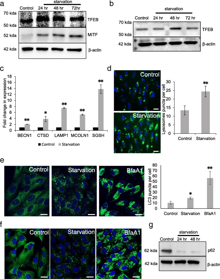
Methods: Expression of TFEB and MITF protein levels were evaluated in cells subjected to prolonged periods of nutrient deprivation. mRNA levels of the CLEAR network genes was measured by quantitative real time PCR (qRT-PCR) analysis in cells deprived of nutrients, treated with ammonium chloride and upon overexpression of constitutively active TFEB. Immunostaining with LC3 antibody was used to measure autophagy flux. Labeling with lysoTracker dye was used to assess lysosomes.

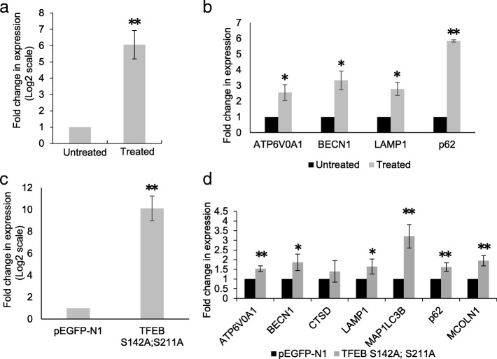
Results: Our results show that nutrient deprivation increases protein levels of TFEB and MITF in ARPE-19 cells. Nutrient stress induces the expression of lysosomal (LAMP1, CTSD MCOLN1, SGSH) and autophagy (BECN1) genes. Lysosomal stress also increases the expression of lysosomal (ATP6V0A1 and LAMP1) and autophagy (p62 and BECN1) genes. Our results show that overexpression of constitutively active TFEB also induces the expression of CLEAR network genes.

Conclusions: Collectively, these observations suggest that nutrient stress induces the protein expression of both MITF and TFEB in ARPE-19 cells. TFEB-regulated transcriptional program plays an important role in adaptive response of cells during both nutrient and lysosomal stress.

Keywords: Autophagy; Lysosomal stress; Nutrient deprivation.


Conflict of interest statement

Competing interestsThe authors declare that they have no competing interests.

Figures

Fig. 1
Fig. 1
Induction of TFEB and CLEAR network genes in cells subjected to starvation. a ARPE-19 cells were subjected to 24–72 h period of nutrient deprivation and the expression levels of TFEB and MITF was measured by immunoblotting. b Expression of TFEB in RPE choroid extracts from mice subjected to 24–72 h period of nutrient withdrawal. c Quantitative real time PCR (qRT-PCR) analysis was performed to analyze the expression of autophagy and lysosomal genes: BECN1, CTSD, LAMP1, MCOLN1 and SGSH in ARPE-19 cells subjected to nutrient deprivation for 48 h. d LysoTracker staining of ARPE-19 cells in cells subjected to nutrient deprivation for 24 h. e ARPE-19 cells were subjected to a 24 h period of nutrient deprivation to determine the cellular expression of LC3 by immunostaining. f Immunostaining with p62 antibody to determine cellular levels of p62 in cells subjected to nutrient deprivation and Bafilomycin treatment for 24 h. g Immunoblot analysis to determine the expression of p62 in cells subjected to nutrient deprivation for 24 and 48 h. Values represent mean ± s.d. of three independent experiments. For animal experiments n = 3 mice were used per group. Student’s t-test (two tailed) was used. For quantification of images Mann–Whitney U test was used. *P-value <0.05 and **P-value <0.01. Scale = 20 μm
Fig. 2
Fig. 2
Transcriptional induction of TFEB and CLEAR network genes in cells subjected to treatment with ammonium chloride and upon overexpression of TFEB. a Expression levels of TFEB transcripts in ARPE-19 cells treated with ammonium chloride. b The expression of autophagy and lysosomal genes: ATP6V0A1, BECN1, LAMP1 and p62was analyzed by qRT-PCR in cells treated with ammonium chloride. c ARPE-19 cells were transfected with a constitutively active mutant of TFEB (S142A; S211A) and the cellular levels of TFEB transcripts was analyzed by qRT-PCR. d qRT-PCR analysis of the expression of lysosomal (LAMP-1, CTSD, MCOLN1 and ATP6V0A1) and autophagy genes (BECN1, MAP1LC3B and p62) upon overexpression of constitutively active TFEB in ARPE-19 cells. Values represent mean ± s.d. of three independent experiments. Student’s t-test (two tailed) was used for analysis *P-value <0.05; **P-value <0.01

Similar articles

Cited by

References

    1. Boulton M, Dayhaw-Barker P. The role of the retinal pigment epithelium: topographical variation and ageing changes. Eye. 2001;15:384. doi: 10.1038/eye.2001.141. - DOI - PubMed
    1. Mazzoni F, Safa H, Finnemann SC. Understanding photoreceptor outer segment phagocytosis: use and utility of RPE cells in culture. Exp Eye Res. 2014;126:51–60. doi: 10.1016/j.exer.2014.01.010. - DOI - PMC - PubMed
    1. Kevany BM, Palczewski K. Phagocytosis of retinal rod and cone photoreceptors. Physiology (Bethesda, Md) 2010;25(1):8–15. - PMC - PubMed
    1. Bosch E, Horwitz J, Bok D. Phagocytosis of outer segments by retinal pigment epithelium: phagosome-lysosome interaction. J Histochem Cytochem. 1993;41(2):253–263. doi: 10.1177/41.2.8419462. - DOI - PubMed
    1. Chen H, Lukas TJ, Du N, Suyeoka G, Neufeld AH. Dysfunction of the retinal pigment epithelium with age: increased Iron decreases phagocytosis and lysosomal activity. Invest Ophthalmol Vis Sci. 2009;50(4):1895–1902. doi: 10.1167/iovs.08-2850. - DOI - PubMed

MeSH terms

Substances