Review
. 2019 Apr;8(2):114-126.
doi: 10.21037/tp.2019.04.06.

Acute therapy of newborns with critical congenital heart disease

Affiliations
Review

Acute therapy of newborns with critical congenital heart disease

Markus Khalil et al. Transl Pediatr. 2019 Apr.

Abstract

Critical congenital heart disease (cCHD) is the most common reason for acute cardiac failure in the neonatal period. cCHD, defined by systemic low cardiac output (LCO) and requiring surgery or catheter-based intervention in the first year of life, has an incidence of approximately 15% of CHD and is responsible for up to 25% fatalities of newborn infants. Clinical deterioration develops in most cases due to rapid closure of the ductus arteriosus (DA). Early diagnosis and immediate treatment determinate beneficial outcome. Critical CHD can be classified in duct-dependent systemic flow, duct-dependent pulmonary flow and transposition of the great arteries. The latter two manifest themselves in oxygen resistant cyanosis, whereas CHD with duct-dependent systemic flow may present itself with cardiogenic shock, which can be difficult to differentiate from other causes of shock such as sepsis. Besides prostaglandin therapy for reopening the arterial duct, a balanced parallel pulmonary and systemic circulation should be a therapeutic goal. In CHD with duct-dependent systemic flow a decrease of pulmonary resistance should be avoided; therefore inadequate oxygen therapy, hyperventilation and alkalosis due to excessive treatment of acidosis, should be averted. Volume therapy should be performed carefully. In CHD with duct-dependent pulmonary flow, pulmonary resistance can be decreased, in case of poor pulmonary flow systemic resistance should be increased, mild alkalosis is recommended. Intense volume therapy is in most cases necessary, except if a restrictive atrial communication is present. In addition to intensive care measures, an arsenal of catheter- and surgery-based procedures need to be hold available as back-up for emergency procedures. Transcatheter interventions are nowadays decisive. Atrial-septostomy was the first and still the most utilized high-urgency procedure; DA-stenting is used in prostaglandin-refractory duct stenosis. In the presence of critical aortic valve stenosis, palliation consists of balloon valvuloplasty. In critical aortic coarctation with myocardial failure and no response to prostaglandin, palliative balloon angioplasty may be the method of choice as bridging for corrective surgery.

Keywords: Newborns; critical congenital heart disease (cCHD); fetal circulation; postnatal management; transcatheter therapy.


Conflict of interest statement

Conflicts of Interest: The authors have no conflicts of interest to declare.

Figures
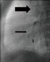
Figure 1
Figure 1
Newborn with hypoplastic left heart treated by Giessen hybrid approach. Lateral 90°: placing of a self-expandable 8 mm × 20 mm Sinus SuperFlex stent (OptiMed, Ettlingen, Germany) within the arterial duct (broad arrow), and an 8 mm × 18 mm Sinus SuperFlex stent within the atrial septum (slender arrow).
Figure 2
Figure 2
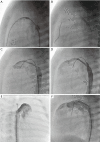
Patient with HLHC and stenotic PDA. (A) Lateral 90°: PDA, almost closed, is crossed with a soft coronary wire from the aortic side; (B) lateral 90°: balloon dilatation of the stenotic PDA with an angioplasty balloon (3.5 mm × 15 mm Trek, Abbott); (C) lateral 90°: delineation of the PDA after balloon angioplasty reveals still a stenotic PDA. (D) Lateral 90°: after crossing of the PDA from the venous side, a balloon-expandable stent (Formula 6 mm × 16 mm, Cook) is brought into position. A wire and a catheter in the aorta serve as marker of the aortic duct junction and therefore enable exact positioning of the stent before inflation; (E) RAO 30°: is in most cases the best angle for visualizing the aortic duct junction. Wire in the aortic arch shows that the stent covers just the PDA and is not protruding into the aorta; (F) lateral 90°: implanted stent with now patent PDA. HLHC, hypoplastic left heart complex; PDA, persistent ductus arteriosus; RAO, right anterior oblique.
Figure 3
Figure 3
Patient with aortic valvular stenosis. (A) Aortic angiography depicts doming of the aortic valve, diameter of the valvular hinges is measurable. Note the angiographic jet, which can serve as guide for crossing of the wire. A pacing catheter is placed in the RV for rapid pacing; (B) balloon valvuloplasty with 6 mm × 20 mm low-pressure balloon under rapid pacing (Tyshak Balloon, PFM, Cologne, Germany).
Figure 4
Figure 4
A 5-day-old newborn with critical coarctation and shock. (A) RAO 20°; (B) lateral 90°: crossing of the coarctation with a 4 F multipurpose catheter and soft coronary wire. Angiography reveals a severe circumscript coarctation; (C) balloon angioplasty with a 6 mm × 20 mm Sterling Balloon (Boston Scientific, Marlborough, USA); (D) aortography after Balloon angioplasty shows a significant wider stenotic segment. RAO, right anterior oblique.

References

    1. Mitchell SC, Korones SB, Berendes HW. Congenital heart disease in 56,109 births. Incidence and natural history. Circulation 1971;43:323-32. 10.1161/01.CIR.43.3.323 - DOI - PubMed
    1. Hoffman JI, Kaplan S. The incidence of congenital heart disease. J Am Coll Cardiol 2002;39:1890-900. 10.1016/S0735-1097(02)01886-7 - DOI - PubMed
    1. Dolk H, Loane M, Garne E. Congenital heart defects in Europe: prevalence and perinatal mortality, 2000 to 2005. Circulation 2011;123:841-9. 10.1161/CIRCULATIONAHA.110.958405 - DOI - PubMed
    1. Oster ME, Lee KA, Honein MA, et al. Temporal trends in survival among infants with critical congenital heart defects. Pediatrics 2013;131:e1502-8. 10.1542/peds.2012-3435 - DOI - PMC - PubMed
    1. Oster ME, Kim CH, Kusano AS, et al. A population-based study of the association of prenatal diagnosis with survival rate for infants with congenital heart defects. Am J Cardiol 2014;113:1036-40. 10.1016/j.amjcard.2013.11.066 - DOI - PMC - PubMed