Review
. 2020 Apr;139(4):643-665.
doi: 10.1007/s00401-019-02029-5. Epub 2019 Jun 4.

An update on the CNS manifestations of neurofibromatosis type 2

Affiliations
Review

An update on the CNS manifestations of neurofibromatosis type 2

Shannon Coy et al. Acta Neuropathol. 2020 Apr.

Erratum in

Abstract

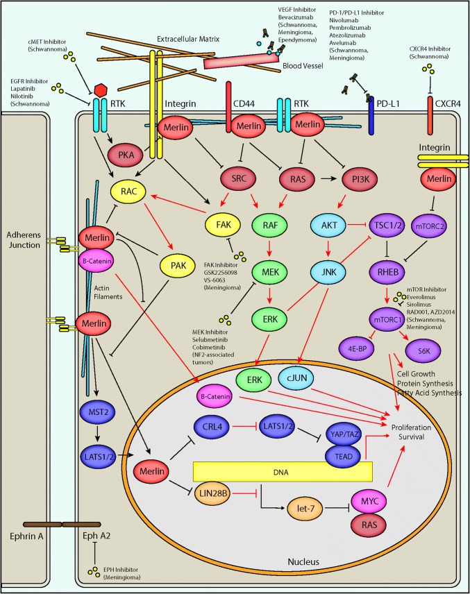
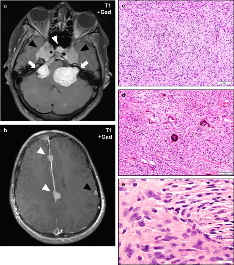
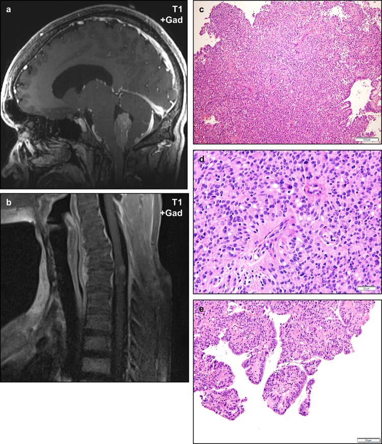
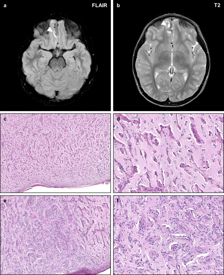
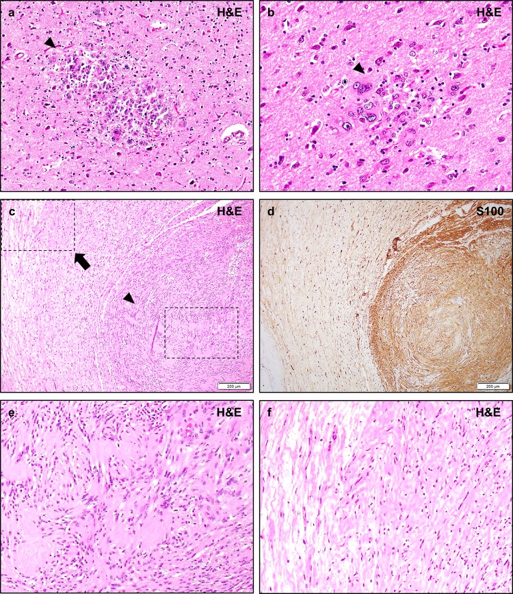
Neurofibromatosis type II (NF2) is a tumor predisposition syndrome characterized by the development of distinctive nervous system lesions. NF2 results from loss-of-function alterations in the NF2 gene on chromosome 22, with resultant dysfunction of its protein product merlin. NF2 is most commonly associated with the development of bilateral vestibular schwannomas; however, patients also have a predisposition to development of other tumors including meningiomas, ependymomas, and peripheral, spinal, and cranial nerve schwannomas. Patients may also develop other characteristic manifestations such as ocular lesions, neuropathies, meningioangiomatosis, and glial hamartia. NF2 has a highly variable clinical course, with some patients exhibiting a severe phenotype and development of multiple tumors at an early age, while others may be nearly asymptomatic throughout their lifetime. Despite the high morbidity associated with NF2 in severe cases, management of NF2-associated lesions primarily consists of surgical resection and treatment of symptoms, and there are currently no FDA-approved systemic therapies that address the underlying biology of the syndrome. Refinements to the diagnostic criteria of NF2 have been proposed over time due to increasing understanding of clinical and molecular data. Large-population studies have demonstrated that some features such as the development of gliomas and neurofibromas, currently included as diagnostic criteria, may require further clarification and modification. Meanwhile, burgeoning insights into the molecular biology of NF2 have shed light on the etiology and highly variable severity of the disease and suggested numerous putative molecular targets for therapeutic intervention. Here, we review the clinicopathologic features of NF2, current understanding of the molecular biology of NF2, particularly with regard to central nervous system lesions, ongoing therapeutic studies, and avenues for further research.

Keywords: Acoustic neuroma; Cellular schwannoma; Central neurofibromatosis; ERM (ezrin/radixin/moesin) family scaffolding; Ependymoma; Epidemiology of familial tumor syndromes; Gardner; Glial hamartia; Glial micro-hamartoma; Glioma; Intraneural schwannoma; LZTR1; Manchester (NIH) Criteria; Manchester Criteria; Meningioangiomatosis; Meningioma; Merlin; NF2; Neurofibroma; Neurofibromatosis type 2; Neurofibromatosis type II; Neurofibromin 2; Plexiform schwannoma; Posterior subcapsular lenticular opacities; SH3PXD2A-HTRA1; SMARCB1; SMARCE1; SUFU; Schwannoma; Schwannomatosis; Schwannomin; Verocay body; Vestibular schwannoma; Von Recklinghausen; Wishart.


Conflict of interest statement

The authors declare no conflicts of interest.

Figures

Fig. 1
Fig. 1
NF2/merlin signaling and potential therapeutic targets in NF2. A schematic diagram depicts major intracellular pathways regulated by merlin, the protein product of the NF2 gene. Merlin is associated with the plasma membrane via its N-terminal FERM domain, where it interacts with adherens junctions and numerous cell surface receptors including integrins, receptor tyrosine kinases (RTK), and CD44. The C-terminal domain of merlin interacts with cytoskeletal actin filaments. Merlin has a predominantly inhibitory activity on downstream effectors including RAS, PI3K, RAC, and SRC, which may result in reduced downstream RAF/MEF/ERK, AKT/JNK/JUN, mTORC1, FAK, and RAC signaling. Merlin may inhibit translocation of β-catenin to the nucleus, reducing the output of canonical Wnt/β-catenin signaling. Merlin also interacts with the Hippo pathway by promoting MST1/2-mediated translocation of LATS1/2 to the nucleus as well as through inhibition of CRL4DCAF1, resulting in reduced transcriptional output of YAP/TAZ and TEA domain transcription factors (TEADs). In the nucleus, merlin may also inhibit the activity of LIN28A, thereby relieving inhibition of the let-7 miRNA cluster leading to downregulation of proto-oncogenic proteins such as MYC and RAS. Loss of function of merlin via pathogenic mutations in NF2 patients results in alteration of downstream activity in each of these pathways (red arrows indicate steps with possible increased activity following loss of merlin function, as in NF2-associated tumors), potentially leading to increased cell growth, protein and fatty acid synthesis, proliferation, and survival. A variety of therapeutic avenues have been explored in NF2 patients, including inhibition of proteins regulated by merlin as well as other cellular receptors. Inhibition of the RAF/MEK/ERK pathway via small molecule MEK inhibitors is currently being explored in clinical trials for NF2-associated tumors (NCT02639546, NCT03095248). A phase 2 clinical trial is exploring small molecule inhibition of FAK by GSK2256098 (NCT02523014/A071401) in NF2 mutant meningiomas. Small molecule mTOR inhibitors including everolimus and sirolimus have shown therapeutic promise in NF2-associated schwannomas and meningiomas, and additional compounds including RAD001 and AZD2014 are in clinical trials for the treatment of progressive/symptomatic NF2-associated meningiomas (NCT02831257) and recurrent high-grade (WHO grade II/III) meningiomas (NCT03071874), respectively. The VEGF inhibitor bevacizumab has shown clinical utility in NF2-associated schwannomas, meningiomas, and ependymomas. Inhibition of the tyrosine kinase receptors cMET and EGFR may provide additional therapeutic opportunities in NF2-associated meningiomas. Additional therapeutic targets have been proposed, including the immunomodulatory PD-1/PD-L1 axis, the chemokine receptor CXCR4, and Ephrin receptor A2 (EphA2)
Fig. 2
Fig. 2
Vestibular Schwannomas in NF2. Magnetic resonance imaging (MRI) of an NF2 patient with bilateral vestibular schwannomas involving the cerebellopontine angle and internal acoustic meatus (ad, arrowheads). Vestibular schwannomas are typically isointense on T1-weighted sequences (a), and show heterogeneous signal on T2-weighted sequences (b). Schwannomas typically exhibit intense gadolinium contrast enhancement (cd). Histologically, schwannomas consist of spindled cells with tapering ends and eosinophilic to clear cytoplasm which may be architecturally arranged as densely cellular Antoni A regions with focally palisading nuclei that may form alternating layered hypercellular and eosinophilic paucicellular regions known as Verocay bodies (e, arrowheads), or less cellular Antoni B regions with more prominent extracellular matrix which may have a collagenized or myxoid appearance (f). Varying hypercellular and hypocellular regions may correlate with the heterogeneous intensity of schwannomas on T2-weighted sequences (b). Perivascular hyalinization is often a prominent histologic feature in schwannomas (g, arrowhead) and may aid in the diagnosis of lesions with an unusual histologic appearance. Schwannomas frequently exhibit degenerative changes (also known as “ancient change”) including increased pleomorphism, bizarre nuclei, and hyperchromasia (h). Such changes are not known to be associated with an increased risk of recurrence or malignant transformation. Schwannomas exhibit intense and diffuse cytoplasmic and nuclear staining with the S100 antigen (i). Scale bars 20 μm (e, g, h), 50 μm (f, i). A web-interactive tool for viewing images of NF2-related tumors is also available: http://tumoratlas.org/coy-acta-neuropathol-2019
Fig. 3
Fig. 3
Non-vestibular schwannomas in NF2. Magnetic resonance imaging (MRI) of an NF2 patient with numerous intracranial and peripheral schwannomas. Coronal post-contrast T1-weighted sequences show bilateral vestibular (CNVIII) schwannomas (arrowheads) confirming the diagnosis of NF2, as well as bilateral trigeminal (CNV) nerve masses (large arrows), bilateral cervical nerve root masses (small arrows), and a post-auricular mass (asterisk) consistent with schwannomas (a). This patient also developed oculomotor (CNIII) schwannomas (b, arrowhead), and a small left trochlear nerve (CNIV) schwannoma (c, arrowhead). Whole body inversion recovery (IR) sequences showed large plexiform masses in the bilateral brachial plexus (d, arrowheads), and right sacral plexus (e, arrowhead). Axial slices of the latter mass demonstrate the multi-nodular plexiform architecture of the lesion (f, arrowhead). Schwannomas may also arise in unusual locations, as illustrated by a cutaneous lesion in the scalp (g, arrowhead), intramuscular lesion in the right iliopsoas muscle (h), and a diffusely infiltrative lesion in the right Achilles tendon (i, arrowhead) in this patient
Fig. 4
Fig. 4
Histologic appearance of non-vestibular schwannomas in NF2. Non-vestibular peripheral schwannomas in NF2 patients may exhibit unusual histologic appearances. Skin biopsy of a cutaneous plaque-like plexiform schwannoma in an NF2 patient shows a nodular and infiltrative proliferation of neoplastic cells involving the dermis and subcutis (a). While sporadic schwannomas are typically circumscribed lesions with a pushing growth pattern, cutaneous schwannomas may infiltrate around skin adnexal structures such as hair follicles and sebaceous glands (b), and deeper eccrine glands and ducts (c). A plexiform schwannoma in the soft tissue of an NF2 patient shows multi-nodular growth along peripheral nerves (d), with typical morphologic features including palisading nuclei and Verocay bodies (e, f). NF2 patients may also develop intraneural schwannomas (g) with whorls of neoplastic cells infiltrating between individual nerve fibers, highlighted by S100 (h) and neurofilament protein (NFP) (i, j) immunohistochemistry. Schwann cell tumorlets may be identified in spinal nerve roots and peripheral nerves, and are hypothesized to represent precursor lesions to larger solitary or plexiform schwannomas (k, l). Scale bars 200 μm (a, d, g, h, i, k), 100 μm (c), 50 μm (b, e, j), 10 μm (f)
Fig. 5
Fig. 5
Meningiomas in NF2. NF2 patients frequently develop multiple meningiomas, which may involve unusual anatomic locations. Post-gadolinium T1-weight magnetic resonance imaging of the skull base of an NF2 patient with bilateral vestibular schwannomas (a, arrows) and durally based masses consistent with meningiomas in the bilateral Meckel’s caves (black arrowheads) and cavernous sinus (white arrowhead). This patient also had multiple meningiomas involving the falx cerebri (white arrowheads) and cerebral convexity (black arrowhead) (b). Histologically, NF2-associated meningiomas most often exhibit fibrous morphology (c), though any histologic pattern may be encountered. A second NF2-associated meningioma shows scattered psammoma bodies (d). The cells of NF2-associated schwannomas are cytologically similar to sporadic meningiomas and most often exhibit ovoid to spindled cells with find chromatin, scattered nuclear pseudo-inclusions, and mild cytologic atypia (e). Scale bars 50 μm (c, d), 10 μm (e)
Fig. 6
Fig. 6
Ependymomas in NF2. Ependymomas are frequently diagnosed in NF2 patients and may occur in a variety of locations including unusual sites. Post-gadolinium T1-weighted magnetic resonance imaging of an NF2 patient shows a weakly enhancing intraventricular mass in the fourth ventricle (a). A second NF2 patient developed an intramedullary mass in the cervical spine (b). Histologically, the fourth ventricular mass exhibited typical features of ependymoma including prominent perivascular pseudorosettes and scattered true ependymal rosettes (c, d), with some regions exhibiting prominent papillary architecture (e). Scale bars 100 μm (c), 20 μm (d), 50 μm (e)
Fig. 7
Fig. 7
NF2-associated meningioangiomatosis. Meningioangiomatosis is a rare plaque-like leptomeningeal and perivascular proliferation of fibroblastic and meningothelial-appearing cells which may extend along Virchow–Robin spaces. Such lesions are typically hyperintense on FLAIR (a) and T2-weighted sequences (b), with intense contrast enhancement on post-gadolinium T1-weighted sequences. Histologic examination of this lesion from an NF2 patient with refractory epilepsy shows a mixed perivascular proliferation of fibrovascular (c, d) or meningothelial-like cells (e, f) extending from the pial surface into the cortical parenchyma. The intervening cortex shows intact neurons with no definite evidence of focal cortical dysplasia. The neoplastic cells are typically well differentiated with no necrosis, atypia, or prominent nucleoli
Fig. 8
Fig. 8
Other lesions in NF2. Post-mortem examination of NF2 patients may demonstrate small circumscribed collections of immature-appearing glial cells known as glial hamartia (a, b, arrowheads). The cells of these lesions typically exhibit eosinophilic cytoplasm, pleomorphic nuclei, and occasional multi-nucleation. The cells are strongly S100 positive, with occasional focal GFAP staining, but their precise lineage derivation is not certain. The surrounding cortex may exhibit reactive changes but does not typically exhibit features of focal cortical dysplasia. Hybrid schwannoma/neurofibromas are frequently encountered in NF2 patients (c, d), and typically exhibit nodular regions of schwannoma with typical histologic features such as Verocay bodies and hyalinized vessels, a monomorphic cellular population, and strong/diffuse S100 staining (e, arrowhead), while other regions exhibit lower density regions with mixed cellularity, loose “shredded carrot” collagen, and less prominent S100 staining consistent with a neurofibroma component (f, arrow). While pure neurofibromas are occasionally encountered in NF2 patients, they are typically sporadic solitary masses, in contrast to the multiple plexiform neurofibromas of NF1. Pathologic review shows that many lesions initially diagnosed as neurofibromas in NF2 patients are in fact misdiagnosed hybrid schwannoma/neurofibromas. Scale bars 200 μm (c, d)

References

    1. Louis DN, Ohgaki H, Wiestler OD, Cavenee WK. WHO classification of tumors of the central nervous system. Lyon: IARC/WHO; 2016. pp. 294–303. - PubMed
    1. Riccardi VM. Neurofibromatosis: clinical heterogeneity. Curr Probl Cancer. 1982;8:1–34. - PubMed
    1. Ruggieri M, Praticò AD, Caltabiano R, Polizzi A. Early history of the different forms of neurofibromatosis from ancient Egypt to the British Empire and beyond: first descriptions, medical curiosities, misconceptions, landmarks, and the persons behind the syndromes. Am J Med Genet A. 2018;176(3):515–550. doi: 10.1002/ajmg.a.38486. - DOI - PubMed
    1. Von Recklinghausen F. Ueber die multiplen fibroma der haut und ihre beziehung zu den multiplen neuromen. Berlin: A Hirschwald; 1882. - PubMed
    1. Wishart JH. Case of tumours in the skull, dura mater, and brain. Edinb Med Surg J. 1822;18:393–397. - PMC - PubMed

Publication types

MeSH terms