

Effects of dehydration on echocardiographic diastolic parameters in healthy cats
- PMID: 31161736
- PMCID: PMC6538523
- DOI: 10.4142/jvs.2019.20.e18
Effects of dehydration on echocardiographic diastolic parameters in healthy cats
Abstract
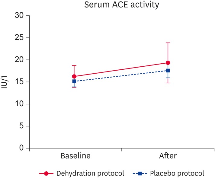
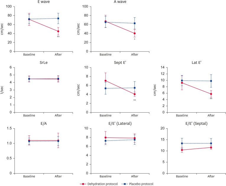
This study aimed to assess the effects of dehydration on echocardiographic indices in healthy cats: specifically, it aimed to assess the effects of volume depletion on diastolic function. Nine experimental cats were subjected to both a dehydration and placebo protocol separated by a 21-day washout period. Echocardiography was performed at baseline and on completion of each protocol. Results were compared between the two protocols. Volume depletion was induced by intravenous administration of furosemide. Volume depletion showed a significant association with increased interventricular septal and left ventricular free wall thickness at end-diastole, decreased left ventricular internal diameter at end-diastole, and left atrial diameter at end-systole. The peak early (E) and late (A) diastolic filling velocities, and the peak early diastolic velocities (E') were significantly decreased by dehydration. Volume depletion did not affect peak longitudinal strain rate during early diastole, E/A, or E/E'. Volume depletion significantly affected the echocardiographic diastolic indices and conventional echocardiographic parameters in healthy cats.
Keywords: Diastolic; feline; furosemide; hypertrophy.
© 2019 The Korean Society of Veterinary Science.
Conflict of interest statement
The authors declare no conflicts of interest.
Figures
Similar articles
-
The effects of maintenance recombinant human erythropoietin therapy on ambulatory blood pressure recordings: conventional, Doppler, and tissue Doppler echocardiographic parameters.Artif Organs. 2005 Dec;29(12):965-72. doi: 10.1111/j.1525-1594.2005.00166.x. Artif Organs. 2005. PMID: 16305652 Clinical Trial.
-
The effect of hydration status on the echocardiographic measurements of normal cats.J Vet Intern Med. 2007 Sep-Oct;21(5):1008-15. doi: 10.1892/0891-6640(2007)21[1008:teohso]2.0.co;2. J Vet Intern Med. 2007. PMID: 17939557
-
[Echocardiographic and Doppler echocardiographic characterization of left ventricular diastolic function].Herz. 1990 Dec;15(6):377-92. Herz. 1990. PMID: 2279732 Review. German.
-
Airflow obstruction and left ventricular filling pressure in suspected chronic obstructive pulmonary disease.Respir Physiol Neurobiol. 2014 Feb 1;192:85-9. doi: 10.1016/j.resp.2013.12.008. Epub 2013 Dec 19. Respir Physiol Neurobiol. 2014. PMID: 24361463
-
[Evaluation of left ventricular diastolic function using Doppler echocardiography].Med Pregl. 1999 Jan-Feb;52(1-2):13-8. Med Pregl. 1999. PMID: 10352498 Review. Croatian.
Cited by
-
ACVIM consensus statement guidelines for the classification, diagnosis, and management of cardiomyopathies in cats.J Vet Intern Med. 2020 May;34(3):1062-1077. doi: 10.1111/jvim.15745. Epub 2020 Apr 3. J Vet Intern Med. 2020. PMID: 32243654 Free PMC article.
-
Case report: Diffuse large B-cell lymphoma presenting as congestive heart failure in a cat.Front Vet Sci. 2024 Sep 25;11:1467448. doi: 10.3389/fvets.2024.1467448. eCollection 2024. Front Vet Sci. 2024. PMID: 39386247 Free PMC article.
-
Case report: A novel occurrence of persistent left cranial vena cava coexisting with polycystic kidney disease in a cat.Front Vet Sci. 2023 Oct 5;10:1268493. doi: 10.3389/fvets.2023.1268493. eCollection 2023. Front Vet Sci. 2023. PMID: 37869489 Free PMC article.
-
The Urinary Hormonal State of Cats Associated With Social Interaction With Humans.Front Vet Sci. 2021 Jul 26;8:680843. doi: 10.3389/fvets.2021.680843. eCollection 2021. Front Vet Sci. 2021. PMID: 34381833 Free PMC article.
References
-
- Campbell FE, Kittleson MD. The effect of hydration status on the echocardiographic measurements of normal cats. J Vet Intern Med. 2007;21:1008–1015. - PubMed
-
- Greene JP, Lefebvre SL, Wang M, Yang M, Lund EM, Polzin DJ. Risk factors associated with the development of chronic kidney disease in cats evaluated at primary care veterinary hospitals. J Am Vet Med Assoc. 2014;244:320–327. - PubMed
-
- Fox PR, Liu SK, Maron BJ. Echocardiographic assessment of spontaneously occurring feline hypertrophic cardiomyopathy. An animal model of human disease. Circulation. 1995;92:2645–2651. - PubMed
Publication types
MeSH terms
Substances
LinkOut - more resources
Full Text Sources
Medical
Miscellaneous
