

doi: 10.1164/rccm.201903-0503LE.
Type I Collagen-targeted Positron Emission Tomography Imaging in Idiopathic Pulmonary Fibrosis: First-in-Human Studies
Affiliations
- PMID: 31161770
- PMCID: PMC6635789
- DOI: 10.1164/rccm.201903-0503LE
Item in Clipboard
Type I Collagen-targeted Positron Emission Tomography Imaging in Idiopathic Pulmonary Fibrosis: First-in-Human Studies
Am J Respir Crit Care Med.
.
No abstract available
Figures
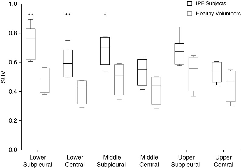
68Ga-CBP8 standardized uptake values (SUVs). Data are reported as box-and-whisker plots. *P < 0.05 and **P < 0.01 for subjects with idiopathic pulmonary fibrosis (IPF) compared with healthy volunteers.
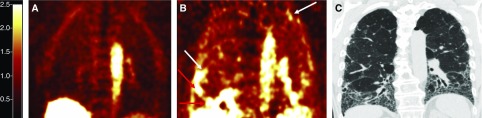
Collagen-targeted molecular imaging using 68Ga-CBP8 positron emission tomography (PET). (A) Coronal PET image of a healthy volunteer, demonstrating low collagen tracer signal in the lungs. (B) PET image of a subject with idiopathic pulmonary fibrosis rendered on the same scale as in A, demonstrating areas of high collagen tracer signal predominantly in subpleural and basilar regions in the lungs. (C) High-resolution computed tomography (CT) of the same subject with idiopathic pulmonary fibrosis obtained within 8 weeks of PET–magnetic resonance imaging. Compared with the PET imaging (B), increased collagen tracer signal is not confined to areas of fibrosis visualized on CT. White arrows highlight regions of high tracer signal within areas of lung without apparent fibrosis on CT. Red arrows highlight regions of high tracer signal within areas of established fibrosis on CT.
References
Publication types
MeSH terms
Substances
Grants and funding
LinkOut - more resources
Full Text Sources
Medical
