

Case Reports
doi: 10.4269/ajtmh.19-0103.
Case Report: Spontaneous Resolution of Intracavitary Hepatic Artery Pseudoaneurysm Caused by Amebic Liver Abscess following Percutaneous Drainage
Affiliations
- PMID: 31162010
- PMCID: PMC6609167
- DOI: 10.4269/ajtmh.19-0103
Item in Clipboard
Case Reports
Case Report: Spontaneous Resolution of Intracavitary Hepatic Artery Pseudoaneurysm Caused by Amebic Liver Abscess following Percutaneous Drainage
Am J Trop Med Hyg.
2019 Jul.
Abstract
Intrahepatic pseudoaneurysm (IHPA) is generally iatrogenic, and it warrants urgent management because of its reportedly significant risk of rupture leading to considerable mortality. Intrahepatic pseudoaneurysm caused by amebic liver abscess (ALA) is exceedingly rare with fewer than seven published reports. All reported symptomatic cases have been treated with hepatic artery embolization; therefore, little is known about its natural course and effect of abscess drainage on its outcome. We report the first case of symptomatic intracavitary IHPA caused by ALA in which ultrasound-guided percutaneous catheter drainage of the abscess resulted in the spontaneous resolution of the IHPA.
Figures
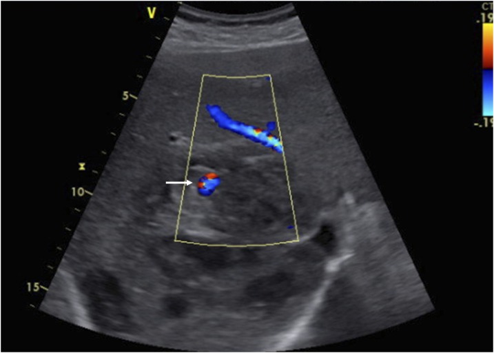
Ultrasonography with color image demonstrates the hypoechoic cystic lesion in the right lobe of the liver representing a partially liquefied amebic liver abscess and an arterial aneurysm demonstrating color flow (arrow). This figure appears in color at www.ajtmh.org .
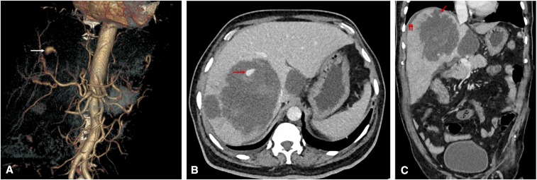
(A) CT angiogram obtained on admission. The volume-rendered image shows an aneurysm of 10-mm size with its feeding artery arising from a branch of the right hepatic artery (arrow). (B) The axial CT image in the venous phase shows multiple hepatic abscesses, including the largest abscess in the right lobe showing a pseudoaneurysm peripherally within the cavity (arrow). The hyperdense areas surrounding the aneurysm represent fresh blood clots. (C) The coronal CT image shows an ill-defined breach in the abscess cavity superiorly (arrow), with small subphrenic fluid collection (double arrow) suggesting a contained rupture. This figure appears in color at www.ajtmh.org .
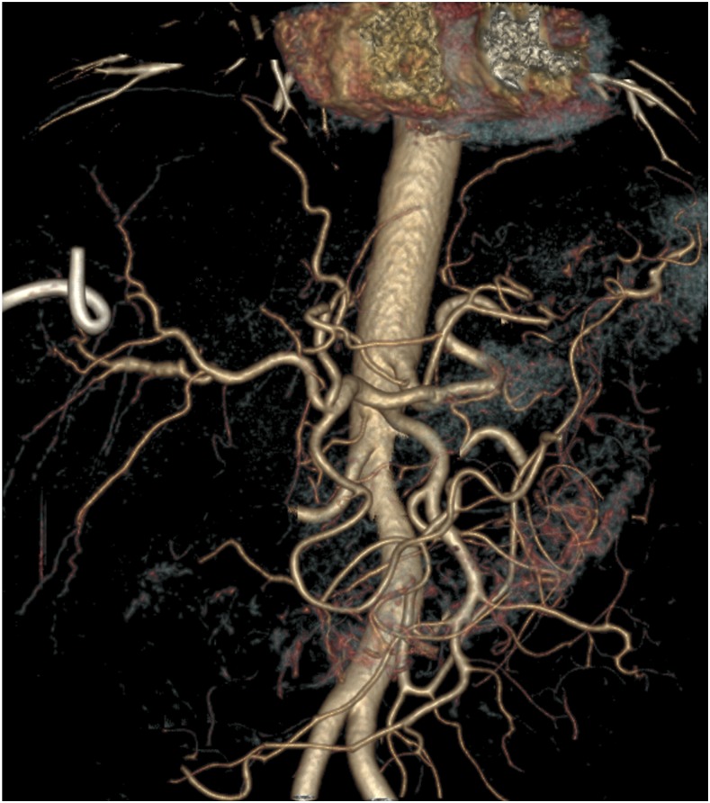
CT angiogram obtained 1 week after the procedure. The volume-rendered image shows complete disappearance of the aneurysm and its feeding artery. This figure appears in color at www.ajtmh.org .
References
-
- Tulsyan N, Kashyap VS, Greenberg RK, Sarac TP, Clair DG, Pierce G, Ouriel K, 2007. The endovascular management of visceral artery aneurysms and pseudoaneurysms. J Vasc Surg 45: 276–283. - PubMed
-
- Berceli SA, 2005. Hepatic and splenic artery aneurysms. Semin Vasc Surg 18: 196–201. - PubMed
-
- Green MH, Duell RM, Johnson CD, Jamieson NV, 2001. Haemobilia. Br J Surg 88: 773–786. - PubMed
-
- Tessier DJ, Fowl RJ, Stone WM, McKusick MA, Abbas MA, Sarr MG, Nagorney DM, Cherry KJ, Gloviczki P, 2003. Iatrogenic hepatic artery pseudoaneurysms: an uncommon complication after hepatic, biliary, and pancreatic procedures. Ann Vasc Surg 17: 663–669. - PubMed
-
- Gopanpallikar A, Rathi P, Sawant P, Gupta R, Dhadphale S, Deshmukh H, 1997. Hepatic artery pseudoaneurysm associated with amebic liver abscess presenting as upper GI hemorrhage. Am J Gastroenterol 92: 1391–1393. - PubMed
Publication types
MeSH terms
LinkOut - more resources
Full Text Sources
