. 2019 Jun 4;5(13):e126241.
doi: 10.1172/jci.insight.126241.

High dietary salt-induced dendritic cell activation underlies microbial dysbiosis-associated hypertension

Affiliations

High dietary salt-induced dendritic cell activation underlies microbial dysbiosis-associated hypertension

Jane F Ferguson et al. JCI Insight. .

Abstract

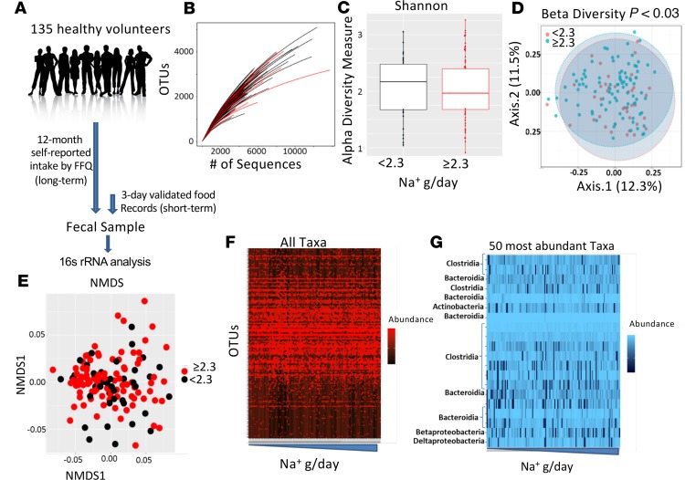
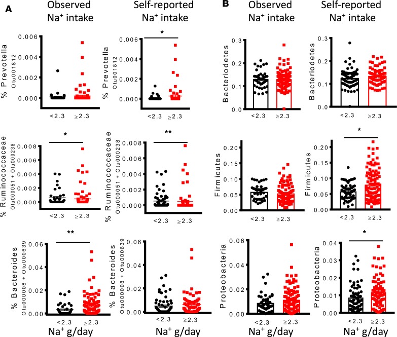
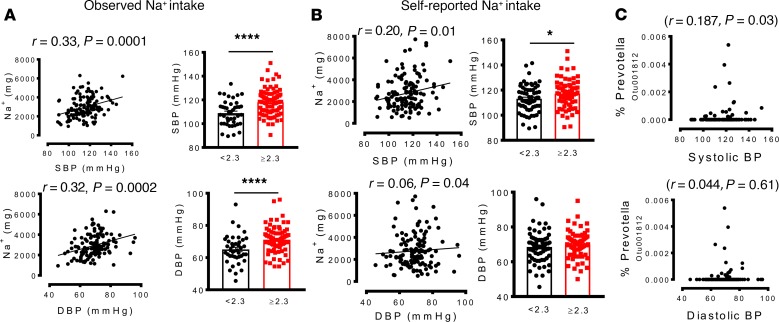
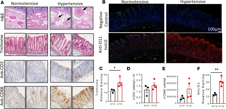
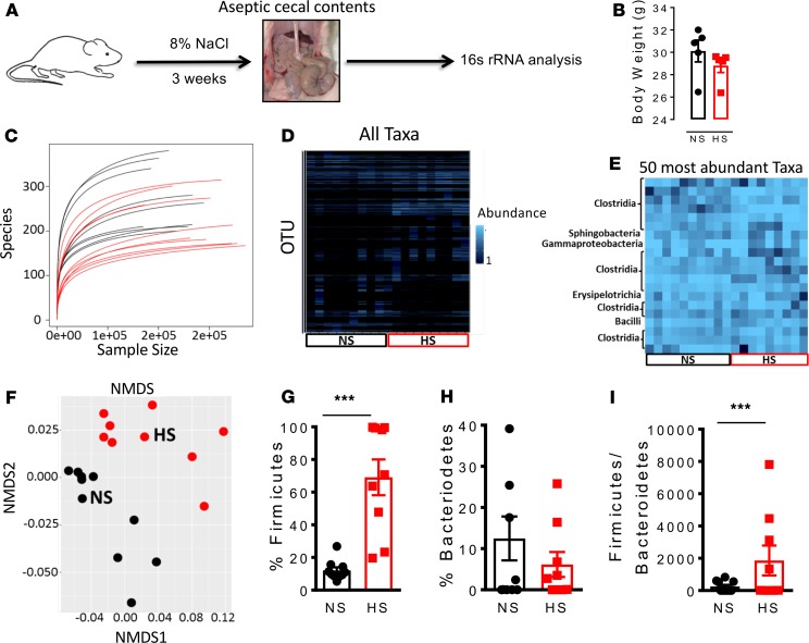
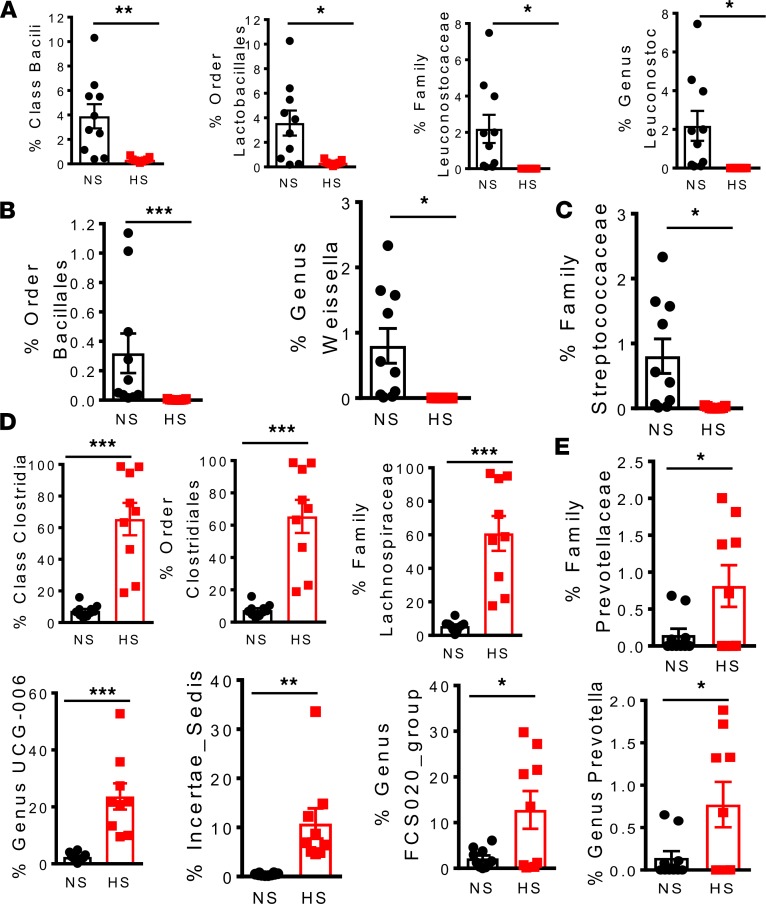
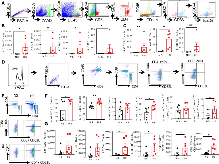
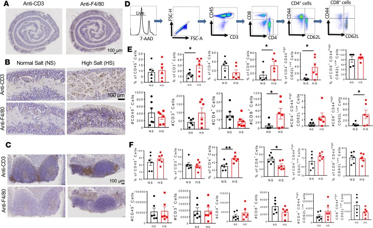
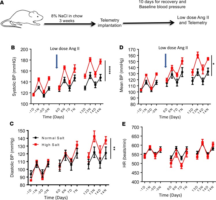
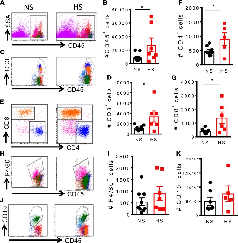
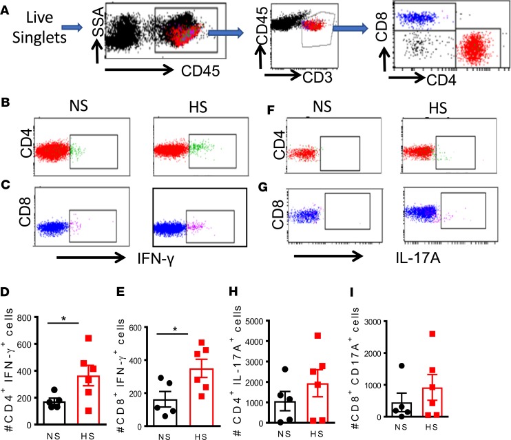
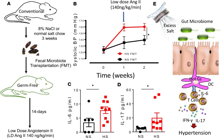
Excess dietary salt contributes to inflammation and hypertension via poorly understood mechanisms. Antigen presenting cells including dendritic cells (DCs) play a key role in regulating intestinal immune homeostasis in part by surveying the gut epithelial surface for pathogens. Previously, we found that highly reactive γ-ketoaldehydes or isolevuglandins (IsoLGs) accumulate in DCs and act as neoantigens, promoting an autoimmune-like state and hypertension. We hypothesized that excess dietary salt alters the gut microbiome leading to hypertension and this is associated with increased immunogenic IsoLG-adduct formation in myeloid antigen presenting cells. To test this hypothesis, we performed fecal microbiome analysis and measured blood pressure of healthy human volunteers with salt intake above or below the American Heart Association recommendations. We also performed 16S rRNA analysis on cecal samples of mice fed normal or high salt diets. In humans and mice, high salt intake was associated with changes in the gut microbiome reflecting an increase in Firmicutes, Proteobacteria and genus Prevotella bacteria. These alterations were associated with higher blood pressure in humans and predisposed mice to vascular inflammation and hypertension in response to a sub-pressor dose of angiotensin II. Mice fed a high salt diet exhibited increased intestinal inflammation including the mesenteric arterial arcade and aorta, with a marked increase in the B7 ligand CD86 and formation of IsoLG-protein adducts in CD11c+ myeloid cells. Adoptive transfer of fecal material from conventionally housed high salt-fed mice to germ-free mice predisposed them to increased intestinal inflammation and hypertension. These findings provide novel insight into the mechanisms underlying inflammation and hypertension associated with excess dietary salt and may lead to interventions targeting the microbiome to prevent and treat this important disease.

Keywords: Hypertension; Inflammation.


Conflict of interest statement

Conflict of interest: The authors have declared that no conflict of interest exists.

Figures

Figure 1
Figure 1. Relationship between a high-salt diet and the human gut microbiome.
(A) Study design; self-reported dietary Na+ intake was estimated from Food Frequency Questionnaires for 12 months (long-term) and 3-day food records prior to the study visit (short-term). 16S rRNA analysis was performed on fecal samples. (B) Rarefaction curve of OTUs in fecal samples. (C) α Diversity measure in people eating a normal salt diet versus a high-salt diet. (D) β Diversity measure in normal versus high salt. (E) Nonmetric multidimensional scaling (NMDS) of bacteria from people eating a low- versus high-salt diet. (F) Relative abundance of selected 16S rRNA gene targets with microbial primers for all taxa. (G) Relative abundance of selected 16S rRNA gene targets with given microbial primers of the 50 most abundant taxa. 16S rRNA data analyses were performed using R software and a type-I error rate of 0.05 was set to infer statistical significance.
Figure 2
Figure 2. A high-salt diet is associated with differences in the human gut microbiome.
Gut microbiome composition in subjects with normal or high-Na+ intake based on American Heart Association recommendations of <2.3 g sodium/d during short-term and long-term intake was analyzed. (A) Relationship between sodium intake and relative abundance of bacterial taxa, including OTUs mapping to the genus Prevotella, family Ruminococcaceae, and genus Bacteroides. (B) Effect of sodium intake on the proportion of Phyla Bacteroidetes, Firmicutes, and Proteobacteria (*P < 0.05, **P < 0.001 using 2-tailed unpaired Student’s t tests).
Figure 3
Figure 3. A high-salt diet is associated with increased colonization of the human gut by genus Prevotella, and this correlates with hypertension.
(A and B) Relationship between sodium intake and systolic and diastolic blood pressures during short-term and long-term intake. (C) Correlation between blood pressure and OTU mapping to Prevotella. (*P < 0.05, ****P < 0.00001 using 2-tailed unpaired Student’s t tests). Linear regression was used to analyze the relationship between blood pressure and Prevotella bacteria, and a type-I error rate of 0.05 was set to infer statistical significance.
Figure 4
Figure 4. Effect of hypertension on gut pathology, inflammation, and isolevuglandin formation in humans.
(A) Representative histological staining images with H&E and Masson’s trichrome for fibrosis as well as immunohistochemical analysis for infiltration of T cells and macrophages of colon sections of normotensive and hypertensive humans obtained from the Vanderbilt Cooperative Human Tissue Network. (B) Immunofluorescent representatives for isolevuglandin (IsoLG) staining. (C) Average data showing trichrome staining in normotensive and hypertensive individuals. (D) Average data for T cells. (E) Average data showing macrophages in hypertensive compared with normotensives. (F) Average data showing IsoLG accumulation in colon sections from hypertensive when compared with normotensive humans (*P < 0.05, **P < 0.001 using 2-tailed unpaired Student’s t tests).
Figure 5
Figure 5. A high-salt diet alters the gut microbiome in mice.
(A) Experimental design; cecal samples from C57BL/6 mice fed a high-salt diet for 3 weeks and regular diet (normal salt) were subjected to 16S rRNA analysis. (B) Body weight of normal salt diet– and high-salt diet–fed mice. (C) Species biodiversity estimation in cecal samples of normal salt diet– and high-salt diet–fed mice. (D) Relative abundance of selected 16S rRNA gene targets with microbial primers for all taxa. (E) Relative abundance of selected 16S rRNA gene targets with given microbial primers of the 50 most abundant taxa. (F) Nonmetric multidimensional scaling (NMDS) showing that the bacteria from normal salt (NS) and high-salt (HS) diet–fed mice cluster separately. Effect of a high-salt diet on the phyla Firmicutes (G) and Bacteroidetes (H). (I) Effect of a high-salt diet on the Firmicutes/Bacteroidetes ratio (***P < 0.0001, n = 10 normal salt and n = 9 high salt using 2-tailed unpaired Student’s t tests).
Figure 6
Figure 6. A high-salt diet depletes lactic acid–producing bacteria and leads to colonization of the gut by families Lachnospiraceae and Prevotellaceae in mice.
(A) Effect of high salt on bacterial number of the Firmicutes phylum belonging to class Bacili, order Lactobacillales, family Leuconostocaceae, and genus Leuconostoc. (B) Effect of high salt on bacterial number of the class Bacilli belonging to the order Bacillales and genus Weissella. (C) Effect of high salt on bacterial numbers of the order Lactobacillales belonging to family Streptococcaceae. (D) Effect of high salt on bacterial number of the Firmicutes phylum belonging to class Clostridia, order Clostridiales, family Lachnospiraceae, and genus Lachnospiraceae UCG-006, Incertae Sedis, and the Lachnospiraceae FCS020 group and (E) family Prevotellaceae and genus Prevotella (*P < 0.05, **P < 0.001, ***P < 0.0001, n = 10 normal salt and n = 9 high salt using 2-tailed unpaired Student’s t tests).
Figure 7
Figure 7. A high-salt diet increases formation of isolevuglandin adducts and inflammation in the mesentery and mesenteric lymph nodes of mice.
(A) Flow cytometry gating strategy to identify inflammatory cell subtypes. (B) Effect of a high-salt diet on mesenteric total CD45+ cells, CD3+ T cells, CD4+ T cells, and CD8+ T cells. (C) Effect of high salt on CD11c+ cells, surface expression of B7 ligand CD86 and intracellular isolevuglandin formation in CD11c+ cells. (D) Flow cytometry gating strategy to identify effector memory T cell subtypes. (E) Representative flow cytometry dot plots for T cell and effector memory T cell subtypes. (F and G) Effect of high-salt feeding on total CD45+ cells, CD3+ T cells, CD4+ T cell, and CD8+ T cells and effector memory CD4+ and CD8+ T cells in the mesenteric lymph nodes (percentages and numbers are shown; *P < 0.05, **P < 0.001 using 2-tailed unpaired Student’s t tests or Mann-Whitney according to the distribution).
Figure 8
Figure 8. A high-salt diet increases inflammation in the colon and Peyer’s patches of mice.
(A) Immunohistochemistry images showing distribution of T cells and monocyte/macrophages in the entire gut of a high-salt diet–fed mouse. Scale bar: 100 μm. (B) Representative immunohistochemistry images showing increased infiltration of T cells and monocyte/macrophages in the colon of high-salt diet–fed mice compared with normal salt diet–fed mice. Scale bar: 100 μm. (C) Representative images showing increased infiltration of T cells and monocyte/macrophages in the Peyer’s patches of high-salt diet–fed mice compared with normal salt diet–fed mice. Scale bar: 100 μm. (D) Flow cytometry gating strategy to identify effector memory T cell subtypes. (E) Effect of high-salt feeding on total CD45+ cells, CD3+ T cells, CD4+ T cells, CD8+ T cells, and effector memory CD4+ and CD8+ T cells in the colon. (F) Effect of high-salt feeding on total CD45+ cells, CD3+ T cells, CD4+ T cells, CD8+ T cells, and effector memory CD4+ and CD8+ T cells in the Peyer’s patches (percentages and numbers are shown; *P < 0.05, **P < 0.001 using 2-tailed unpaired Student’s t tests).
Figure 9
Figure 9. A high-salt diet predisposes to hypertension.
Mice were fed a normal salt diet (black) or a high-salt diet, 8% NaCl (red), for 3 weeks. Osmotic minipumps were implanted at 3 weeks to deliver a subcutaneous low dose of angiotensin II (140 mg/kg/h) for 2 weeks. (A) Experimental strategy. Systolic (B), diastolic (C), mean arterial blood pressure (D), and heart rate (E) were monitored using radiotelemetry (n = 5 normal salt and 6 high salt; *P < 0.05, **P < 0.001, ****P < 0.0001 high salt versus normal salt control, using 2-way repeated-measures ANOVA). D, day; N, night.
Figure 10
Figure 10. A high-salt diet predisposes mice to aortic inflammation in response to low-dose angiotensin II.
Mice were fed a normal salt diet or a high-salt diet, 8% NaCl, for 3 weeks. Osmotic minipumps were implanted at 3 weeks to deliver a subcutaneous low dose of angiotensin II (140 mg/kg/h) for 2 weeks. The mice were sacrificed, and single-cell suspensions were prepared from freshly isolated mouse aortas via enzymatic digestion and mechanical dissociation. Live-cell singlets were analyzed for vascular inflammatory cells including number and percentages CD45+ total leukocytes (A and B), CD3+ T lymphocytes (C and D), CD4+/CD8+T cell subsets (E, F, and G), F4/80+ monocytes and macrophages (H and I), and CD19+ B lymphocytes (J and K). An unpaired t test was used to compare infiltrating leukocyte subsets between normal salt (NS) and high salt (HS) (*P < 0.05 using 2-tailed unpaired Student’s t tests).
Figure 11
Figure 11. Cytokine production of T cells in the spleen of mice fed normal or high-salt diet.
Mice were fed a normal salt diet or a high-salt diet, 8% NaCl, for 3 weeks. Osmotic minipumps were implanted at 3 weeks to deliver a subcutaneous low dose of angiotensin II (140 mg/kg/h) for 2 weeks. The mice were sacrificed, and single-cell suspensions were prepared from freshly isolated mouse spleens via enzymatic digestion and mechanical dissociation. (A) Gating strategy for identifying T cell populations among total splenocytes. Intracellular staining for IFN-γ (BE) and IL-17A (FI) production in T cell subsets (*P < 0.05 using 2-tailed unpaired Student’s t tests). NS, normal salt; HS, high salt.
Figure 12
Figure 12. Fecal microbial transfer from mice fed a high-salt diet predisposes recipient germ-free mice to inflammation and hypertension.
(A) Experimental design for fecal material transfer (FMT) from conventionally fed mice fed a high-salt diet into recipient germ-free mice. (B) Tail cuff systolic blood pressure of normal salt (NS) and high salt (HS) FMT recipient germ-free mice in response to a low subpressor dose of angiotensin II. (C and D) Plasma levels of IL-6 and IL-17 in recipient germ-free mice with FMT from high-salt diet–fed mice when compared with those with FMT from normal salt–fed mice (*P < 0.05 using 2-way repeated-measures ANOVA). (E) Paradigm illustrating how excess dietary salt alters the gut microbiome and activates DCs, leading to hypertension. A high-salt diet compromises the microbiome and induces production of IsoLG protein adduct formation in CD11c+ cells. The DCs become activated and promote T cell production of IL-17 and IFN-γ, leading to hypertension.

References

    1. Whelton PK, et al. 2017 ACC/AHA/AAPA/ABC/ACPM/AGS/APhA/ASH/ASPC/NMA/PCNA Guideline for the Prevention, Detection, Evaluation, and Management of High Blood Pressure in Adults: Executive Summary: A Report of the American College of Cardiology/American Heart Association Task Force on Clinical Practice Guidelines. Hypertension. 2018;71(6):1269–1324. doi: 10.1161/HYP.0000000000000066. - DOI - PubMed
    1. Kearney PM, Whelton M, Reynolds K, Muntner P, Whelton PK, He J. Global burden of hypertension: analysis of worldwide data. Lancet. 2005;365(9455):217–223. doi: 10.1016/S0140-6736(05)17741-1. - DOI - PubMed
    1. Murray CJ, Lopez AD. Measuring the global burden of disease. N Engl J Med. 2013;369(5):448–457. doi: 10.1056/NEJMra1201534. - DOI - PubMed
    1. Lev-Ran A, Porta M. Salt and hypertension: a phylogenetic perspective. Diabetes Metab Res Rev. 2005;21(2):118–131. doi: 10.1002/dmrr.539. - DOI - PubMed
    1. Weinberger MH, Fineberg NS, Fineberg SE, Weinberger M. Salt sensitivity, pulse pressure, and death in normal and hypertensive humans. Hypertension. 2001;37(2 Pt 2):429–432. - PubMed

Publication types

MeSH terms