Review
. 2019 Aug;28(8):756-764.
doi: 10.1097/IJG.0000000000001293.

Gravitational Influence on Intraocular Pressure: Implications for Spaceflight and Disease

Affiliations
Review

Gravitational Influence on Intraocular Pressure: Implications for Spaceflight and Disease

Alex S Huang et al. J Glaucoma. 2019 Aug.

Abstract

Spaceflight-associated neuro-ocular syndrome (SANS) describes a series of morphologic and functional ocular changes in astronauts first reported by Mader and colleagues in 2011. SANS is currently clinically defined by the development of optic disc edema during prolonged exposure to the weightless (microgravity) environment, which currently occurs on International Space Station (ISS). However, as improvements in our understanding of the ocular changes emerge, the definition of SANS is expected to evolve. Other ocular SANS signs that arise during and after ISS missions include hyperopic shifts, globe flattening, choroidal/retinal folds, and cotton wool spots. Over the last 10 years, ~1 in 3 astronauts flying long-duration ISS missions have presented with ≥1 of these ocular findings. Commensurate with research that combines disparate specialties (vision biology and spaceflight medicine), lessons from SANS investigations may also yield insight into ground-based ocular disorders, such as glaucomatous optic neuropathy that may have the potential to lessen the burden of this irreversible cause of vision loss on Earth.


Figures

Figure 1.
Figure 1.
A) Photo of disc edema after spaceflight. This finding defines SANS. B) Ultrasound showing optic nerve sheath dilatations and posterior globe flattening (arrows). C) Photo showing choroidal folds (black arrow) and cotton wool spot (white arrow). D) Total number of US Astronauts tested for each ocular variable (blue) and the number demonstrating the finding through 2017. Optic disc edema defined as Frisèn Scale grade ≥ 1 from postflight fundoscopy images; Choroidal folds visualized on fundoscopy images and/or optical coherence tomography B-scans when available; Globe flattening based on a subjective call from either MRI or ocular ultrasound images; Refractive error shift based on pre to postflight change in cycloplegic refraction ≥0.75 diopters; cotton wool spot observed on fundoscopy images during or after spaceflight. Images adapted from Mader et al., 2011 and 2013,.
Figure 2.
Figure 2.
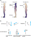
A) Preflight on Earth (1G) arterial blood pressure at the head are approximately 70 mmHg, however the pressure at the feet is 200 mmHg. During microgravity it is hypothesized that arterial blood pressure from head to foot are similar because of the absence of the hydrostatic pressure gradient. Post flight (1G), without the use of countermeasures blood volume is reduced from preflight levels resulting in reduced blood pressure at head level (figure adapted from Zhang and Hargens 2017). B) On Earth, gravity directs body fluid towards the lower extremities in upright positions but toward the head in head-down-tilt position. During weightlessness, in the microgravity environment of space, cephalad fluid shift is seen even in upright positions. Proposed countermeasures theoretically combat this. Lower-extremities cuffs block cephalad fluid flow under weightlessness (blue line with blunted head). Lower-body negative pressure chambers draw fluid to the lower-extremities. Impedance threshold devices draw fluid in the head and into the thoracic cavity. Blue arrows denote overall body fluid flow.
Figure 3.
Figure 3.
A) Photo of Russian Bralset venoconstrictive thigh cuff device. B) Photo of pneumatic venoconstrictive thigh cuff (pVTC) device. C) Photo of mobile venoconstrictive thigh cuff (mVTC) device with modified width and variable pressure adjustment mechanism.
Figure 4.
Figure 4.
Episcleral Veins. Aqueous angiography introduces fluorescent tracers into the eye followed by angiographic imaging in live human subjects, here undergoing routine clinically-indicated cataract surgery. Rows (A-C) show episcleral venous patterns from three eyes from three live patients. Each row is one subject. Segmental patterns are seen showing regions with (green arrows) and without (red arrows) angiographic signal. It is these episcleral veins which may be impacted by body position to alter IOP or be a target for IOP lowering using cytoskeletal relaxing agents. Figure reproduced from (Huang et al., 2018 JOG) with permission from author.

Comment in

References

    1. Mader TH, Gibson CR, Pass AF, et al. Optic disc edema, globe flattening, choroidal folds, and hyperopic shifts observed in astronauts after long-duration space flight. Ophthalmology. 2011;118(10):2058–2069. doi:10.1016/j.ophtha.2011.06.021 - DOI - PubMed
    1. Mader TH, Gibson CR, Otto CA, et al. Persistent Asymmetric Optic Disc Swelling After Long-Duration Space Flight: Implications for Pathogenesis. J Neuro-Ophthalmol Off J North Am Neuro-Ophthalmol Soc. 2017;37(2):133–139. doi:10.1097/WNO.0000000000000467 - DOI - PubMed
    1. Moss HE. Objective Measures of Visual Function in Papilledema. Adv Ophthalmol Optom. 2016;1(1):231–247. doi:10.1016/j.yaoo.2016.03.004 - DOI - PMC - PubMed
    1. Karanjia R, Berezovsky A, Sacai PY, et al. The Photopic Negative Response: An Objective Measure of Retinal Ganglion Cell Function in Patients With Leber’s Hereditary Optic Neuropathy. Invest Ophthalmol Vis Sci. 2017;58(6):BIO300–BIO306. doi:10.1167/iovs.17-21773 - DOI - PubMed
    1. Stenger MB, Tarver WJ, Brunstetter T, et al. NASA Human Research Program Evidence Report: Risk of Spaceflight Associated Neuro-Ocular Syndrome (SANS). NASA Johnson Space Center; 2017. https://humanresearchroadmap.nasa.gov/evidence/reports/SANS.pdf?rnd=0.43.... Accessed February 15, 2018.