. 2019 Aug 7;40(30):2507-2520.
doi: 10.1093/eurheartj/ehz305.

Single-cell transcriptome analyses reveal novel targets modulating cardiac neovascularization by resident endothelial cells following myocardial infarction

Affiliations

Single-cell transcriptome analyses reveal novel targets modulating cardiac neovascularization by resident endothelial cells following myocardial infarction

Ziwen Li et al. Eur Heart J. .

Abstract

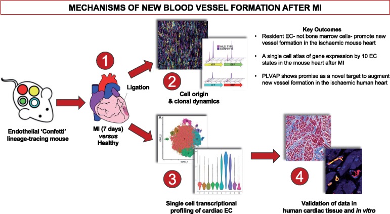
Aims: A better understanding of the pathways that regulate regeneration of the coronary vasculature is of fundamental importance for the advancement of strategies to treat patients with heart disease. Here, we aimed to investigate the origin and clonal dynamics of endothelial cells (ECs) associated with neovascularization in the adult mouse heart following myocardial infarction (MI). Furthermore, we sought to define murine cardiac endothelial heterogeneity and to characterize the transcriptional profiles of pro-angiogenic resident ECs in the adult mouse heart, at single-cell resolution.

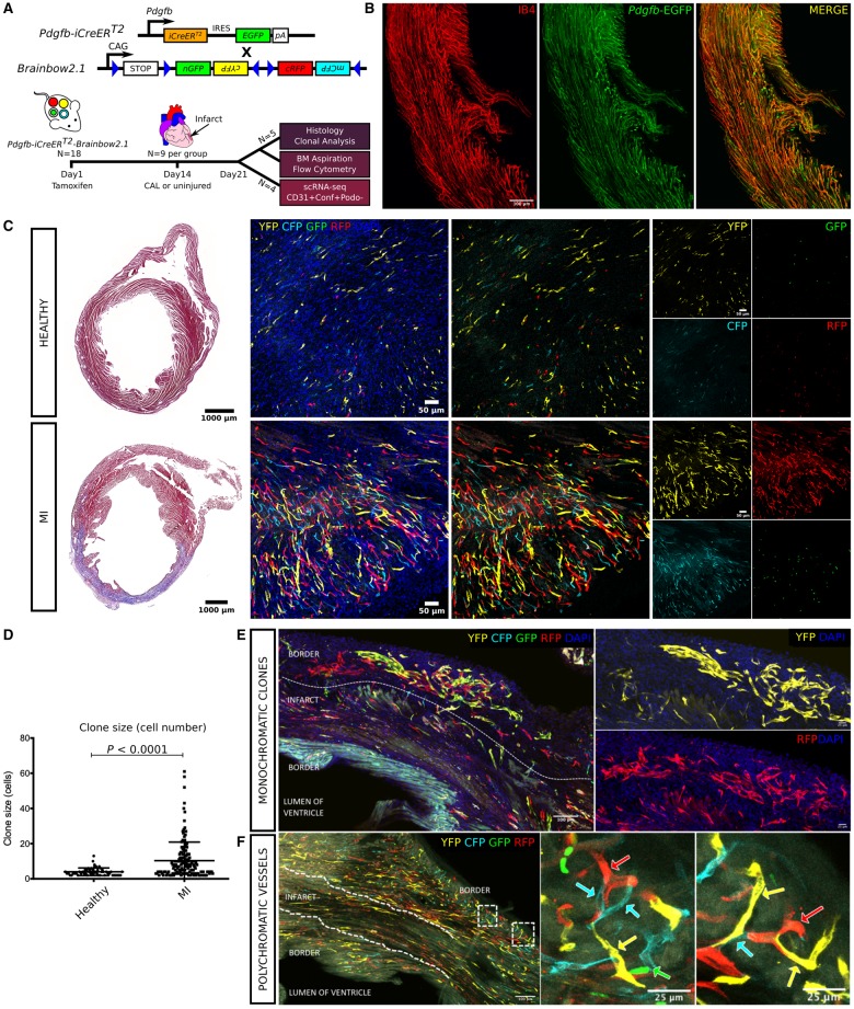
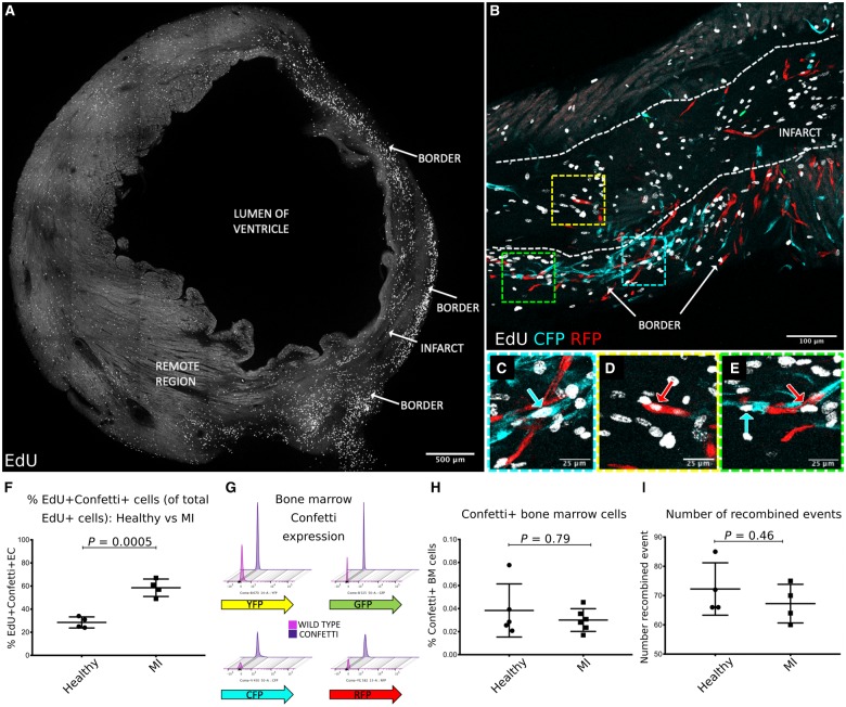
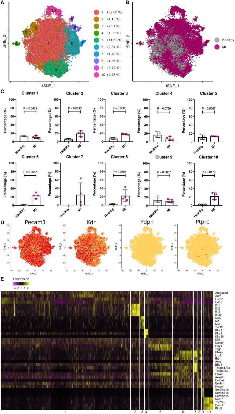
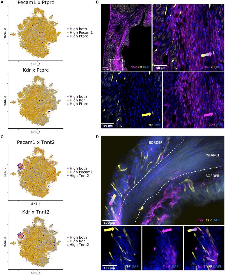
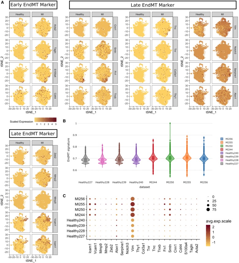
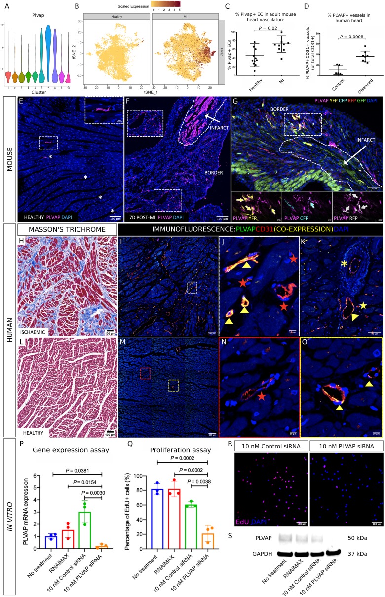
Methods and results: An EC-specific multispectral lineage-tracing mouse (Pdgfb-iCreERT2-R26R-Brainbow2.1) was used to demonstrate that structural integrity of adult cardiac endothelium following MI was maintained through clonal proliferation by resident ECs in the infarct border region, without significant contributions from bone marrow cells or endothelial-to-mesenchymal transition. Ten transcriptionally discrete heterogeneous EC states, as well as the pathways through which each endothelial state is likely to enhance neovasculogenesis and tissue regeneration following ischaemic injury were defined. Plasmalemma vesicle-associated protein (Plvap) was selected for further study, which showed an endothelial-specific and increased expression in both the ischaemic mouse and human heart, and played a direct role in regulating human endothelial proliferation in vitro.

Conclusion: We present a single-cell gene expression atlas of cardiac specific resident ECs, and the transcriptional hierarchy underpinning endogenous vascular repair following MI. These data provide a rich resource that could assist in the development of new therapeutic interventions to augment endogenous myocardial perfusion and enhance regeneration in the injured heart.

Keywords: Cell proliferation; Endothelial cells; Lineage tracing; Myocardial infarction; Single-cell RNA sequencing; Therapeutic angiogenesis.


Figures

Figure 1
Figure 1
Genetic labelling strategy used for study of Pdgfb-iCreERT2 expressing endothelial cells using the R26R-Brainbow2.1 ‘Confetti’ reporter mouse (A). Tamoxifen was administered by intraperitoneal injection followed by coronary artery ligation 14 days later. Injured and healthy hearts) were collected 7 days post-surgery to quantify clonal proliferation and bone marrow reporter expression, or for single-cell RNA sequencing (scRNA-seq) of vascular cardiac Pdgfb-lineage endothelial cells (CD31+ podoplanin Confetti+) (A). Isolectin B4 (IB4) was injected intravenously 15 min prior to cull to identify perfused vessels in the vasculature of Pdgfb-iCreERT2-R26R-Brainbow2.1 adult mouse hearts. Co-expression of isolectin B4 (IB4, red) with Pdgfb-EGFP endothelial cells (green, with co-localized expression shown in orange) demonstrated widespread patency in vessels formed by Pdgfb-lineage endothelial cell clonal proliferation (B). Masson’s trichrome staining confirmed healthy myocardial tissue or the presence of an infarct in each group (C). Clonal proliferation was quantified in 100 μm tissue wholemounts in the infarct border and equivalent healthy region, where a clone was defined as two or more adjacent cells expressing YFP, RFP, nGFP, or mCFP. Clones of each colour were observed throughout the healthy heart with a significant increase in fluorophore expression and clone size in the infarcted (MI) hearts (cells per clone = 4.0 ± 2.1 vs. 10.3 ± 10.6, P <0.0001) (C, D). Clones of >50 cells were commonly observed in the infarct border (E; 2 large multicellular clones in the infarct border expressing RFP or YFP). Vessels with a polychromatic endothelium were observed in healthy and infarcted hearts but were significantly less abundant than monoclonal vessels in both groups (healthy hearts = 25.5 ± 4.1% vs. 74.5 ± 4.1%, P <0.001; MI hearts = 9.3 ± 15.3% vs. 90.7 ± 15.3%, P <0.001) (F; with arrows showing contiguous vessels composed of Pdgfb-lineage endothelial cells expressing different Confetti reporter fluorophores in the infarct border).
Figure 2
Figure 2
A complete transverse section of the left ventricle at 7 days post-MI showing increased density of EdU expressing cells in the border region compared with the infarct and remote region (A). Dense neovascularization due to clonal proliferation of Pdgfb-lineage endothelial cells was observed in the infarct border region (B, with high power inserts in CE). Confetti+Pdgfb-lineage endothelial cells frequently co-expressed EdU (BE) and were significantly increased in the infarct border at 7 days post-MI compared with the Confetti+ EdU+ Pdgfb-lineage endothelial cells in the left ventricle of healthy uninjured mice (28.5 ± 4.8% vs. 58.5 ± 7.6%, P =0.0005) (F). Representative flow cytometry plots showing very low reporter fluorophore expression in femoral bone marrow cells from adult Pdgfb-iCreERT2-R26R-Brainbow2.1 mice are shown in (G) with threshold gates set for each fluorophore using C57Bl6 wild type mice bone marrow cells as a negative control. No change in fluorophore expression by bone marrow cells was observed between healthy and MI groups (0.04 ± 0.02% vs. 0.03 ± 0.009%, P =0.79) (H). The founding number of recombined events (where a Confetti+ cell or clone was counted as one event) was unchanged between healthy and MI groups (72.3 ± 9.0 vs. 67.3 ± 6.6 events per section, P =0.46) (I).
Figure 3
Figure 3
Ten discrete heterogeneous endothelial cell populations were identified in healthy and infarcted adult mouse hearts (A and B). Contributions of cells from both healthy and myocardial infarction hearts to each cluster were compared, and cluster 2, 3, 6, 8, and 10 were largely constituted by the cells from the myocardial infarction hearts. Cluster 7 was composed exclusively of cells from the myocardial infarction hearts (C). Broad expression of endothelial cell markers, such as Pecam1 and Kdr, and the rare presence of Pdpn and Ptprc expressing cells showed purity of the studied endothelial cell population (D). Top differentially expressed marker genes were shown for each of the 10 clusters in the heatmap (E).
Figure 4
Figure 4
Rare Ptprc+ (CD45) cells that co-expressed high levels of Pecam-1 (0.29% total cells) or KDR (0.19% total cells) were dispersed throughout several clusters in the MI group (A). Cells that co-expressed CD45 and a Confetti reporter fluorophore were observed at the infarct border in 100 μm tissue wholemounts (B, arrows). Cluster 3 showed co-expression of endothelial markers, Pecam-1 and Kdr, with genes encoding cardiac muscle markers including troponin-T (Tnnt2) (C). Cells that co-expressed a Brainbow2.1 fluorophore and the cardiomyocyte marker, troponin-T, were identified in the infarct border region in 100 μm tissue wholemounts (D, arrows). These cells were scarce and resembled vascular structures in morphology, rather than cardiomyocytes.
Figure 5
Figure 5
Endothelial-to-mesenchymal transition/partial endothelial-to-mesenchymal transition was investigated in our data using a comprehensive panel of markers of early and late endothelial-to-mesenchymal transition (A). The change in an endothelial-to-mesenchymal transition gene signature between healthy and myocardial infarction groups was minimal and not specific to any cluster (B) despite an increase in some markers (C).
Figure 6
Figure 6
Violin plots showing heightened expression of Plvap in clusters 6, 7, and 8 (A). t-SNE plots highlighting an increased Plvap gene expression in cardiac endothelial cells from the myocardial infarction group compared with the healthy group (B). Plvap was significantly increased in the infarct border region at 7 days post-myocardial infarction, compared with the healthy mouse heart (70.5 ± 19.9% vs. 38.7 ± 28.2%, P =0.002) (C). Plvap expression was also increased in endothelial cells adjacent to regions of fibrosis and scarring in the ischaemic human heart, compared with healthy human hearts (% Plvap+ endothelial cells = 36.9 ± 10.1% vs. 11.1 ± 8.8%, P =0.002) (D). Representative images showing immunofluoresence staining for Plvap accompanying the above data showing the change in expression in cardiac endothelial cells in the healthy (E) and injured Pdgfb-iCreERT2-R26R-Brainbow2.1 mouse heart in the infarct border at 7 days post-myocardial infarction (F), with co-expression of Pdgfb-EC Confetti fluorophore-expressing clones (G with high power inserts and arrows highlighting Plvap+ Confetti+ EC). Consecutive sections from ischaemic and healthy human cardiac tissue were stained using Masson’s Trichrome (H = healthy, L = ischaemic) and by immunofluorescence for PLVAP and CD31 (green and red, respectively, with co-expression in yellow; IK = ischaemic, MO = healthy). PLVAP expression was specific to CD31+ cells and was increased in regions of fibrosis and scarring in the diseased compared with the healthy heart. Plvap+ CD31+ vessels are indicated by yellow arrowheads (J, O), CD31+ Plvap vessels indicated by red stars (J, N). Plvap+ CD31+ EC expression in a venule (yellow arrowhead), arteriole (yellow asterisk) and capillary (yellow star) is shown in (K). siRNA gene silencing of Plvap in human umbilical venous endothelial cells gave a significant reduction at the mRNA level compared with control siRNA (RQ = 3.0 ± 0.9 vs. 0.2 ± 0.2, P =0.003) (P) and was confirmed at the protein level by western blot (S). Proliferation assessed using an EdU incorporation assay was significantly inhibited following Plvap gene silencing compared with control siRNA treatment (% EdU+ human umbilical venous endothelial cells = 60.7 ± 3.9% vs. 21.1 ± 11.0%, P =0.0038; Q, R).
Take home figure
Take home figure
Single cell transcriptome analyses provide a single cell gene expression atlas of resident cardiac endothelial cells that promote neovascularisation following ischaemic injury by undergoing clonal proliferation.
None

Comment in

References

    1. Dunlay SM, Roger VL.. Understanding the epidemic of heart failure: past, present, and future. Curr Heart Fail Rep 2014;11:404–415. - PMC - PubMed
    1. Ponikowski P, Voors AA, Anker SD, Bueno H, Cleland JGF, Coats AJS, Falk V, Gonzalez-Juanatey JR, Harjola VP, Jankowska EA, Jessup M, Linde C, Nihoyannopoulos P, Parissis JT, Pieske B, Riley JP, Rosano GMC, Ruilope LM, Ruschitzka F, Rutten FH, van der Meer P; Group Scientific Document Group. 2016 ESC Guidelines for the diagnosis and treatment of acute and chronic heart failure: the task force for the diagnosis and treatment of acute and chronic heart failure of the European Society of Cardiology (ESC)Developed with the special contribution of the Heart Failure Association (HFA) of the ESC. Eur Heart J 2016;37:2129–2200. - PubMed
    1. Thygesen K, Alpert JS, Jaffe AS, Chaitman BR, Bax JJ, Morrow DA, White HD, Group E.. Fourth universal definition of myocardial infarction (2018). Eur Heart J 2019;40:237–269. - PubMed
    1. Madonna R, Van Laake LW, Davidson SM, Engel FB, Hausenloy DJ, Lecour S, Leor J, Perrino C, Schulz R, Ytrehus K, Landmesser U, Mummery CL, Janssens S, Willerson J, Eschenhagen T, Ferdinandy P, Sluijter JP.. Position Paper of the European Society of Cardiology Working Group Cellular Biology of the Heart: cell-based therapies for myocardial repair and regeneration in ischemic heart disease and heart failure. Eur Heart J 2016;37:1789–1798. - PMC - PubMed
    1. Levy D, Kenchaiah S, Larson MG, Benjamin EJ, Kupka MJ, Ho KK, Murabito JM, Vasan RS.. Long-term trends in the incidence of and survival with heart failure. N Engl J Med 2002;347:1397–1402. - PubMed

Publication types