. 2019 Jun 4;14(6):e0215031.
doi: 10.1371/journal.pone.0215031. eCollection 2019.

Delivery of self-amplifying RNA vaccines in in vitro reconstituted virus-like particles

Affiliations

Delivery of self-amplifying RNA vaccines in in vitro reconstituted virus-like particles

Adam Biddlecome et al. PLoS One. .

Abstract

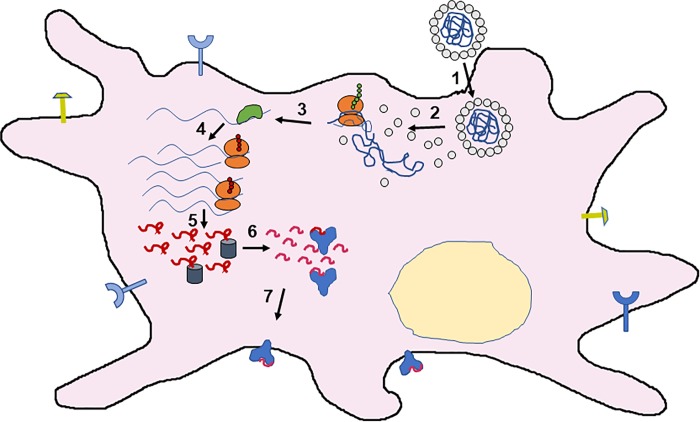
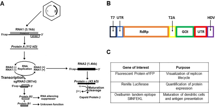
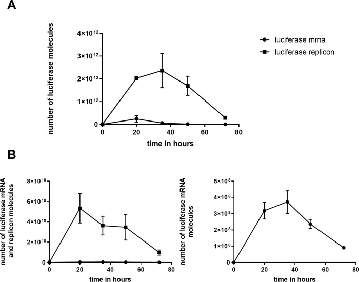
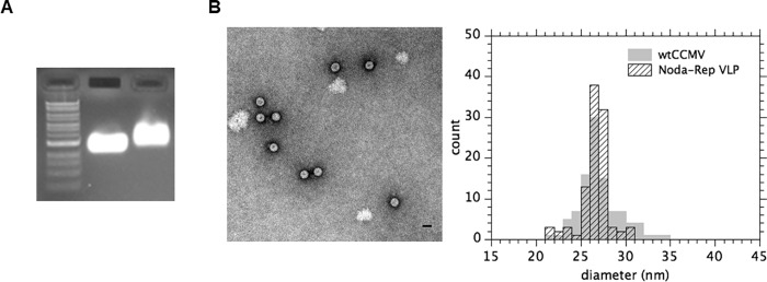
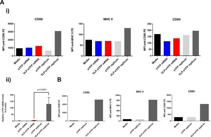
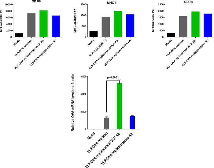
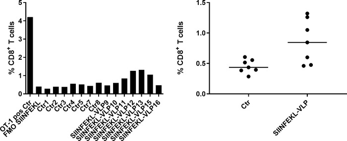
Many mRNA-based vaccines have been investigated for their specific potential to activate dendritic cells (DCs), the highly-specialized antigen-presenting cells of the immune system that play a key role in inducing effective CD4+ and CD8+ T-cell responses. In this paper we report a new vaccine/gene delivery platform that demonstrates the benefits of using a self-amplifying ("replicon") mRNA that is protected in a viral-protein capsid. Purified capsid protein from the plant virus Cowpea Chlorotic Mottle Virus (CCMV) is used to in vitro assemble monodisperse virus-like particles (VLPs) containing reporter proteins (e.g., Luciferase or eYFP) or the tandem-repeat model antigen SIINFEKL in RNA gene form, coupled to the RNA-dependent RNA polymerase from the Nodamura insect virus. Incubation of immature DCs with these VLPs results in increased activation of maturation markers - CD80, CD86 and MHC-II - and enhanced RNA replication levels, relative to incubation with unpackaged replicon mRNA. Higher RNA uptake/replication and enhanced DC activation were detected in a dose-dependent manner when the CCMV-VLPs were pre-incubated with anti-CCMV antibodies. In all experiments the expression of maturation markers correlates with the RNA levels of the DCs. Overall, these studies demonstrate that: VLP protection enhances mRNA uptake by DCs; coupling replicons to the gene of interest increases RNA and protein levels in the cell; and the presence of anti-VLP antibodies enhances mRNA levels and activation of DCs in vitro. Finally, preliminary in vivo experiments involving mouse vaccinations with SIINFEKL-replicon VLPs indicate a small but significant increase in antigen-specific T cells that are doubly positive for IFN and TFN induction.


Conflict of interest statement

The authors have declared that no competing interests exist.

Figures

Fig 1
Fig 1. Schematic of DC activation triggered by VLP delivery of a self-replicating vaccine.
Upon uptake (1), disassembly (2) of the VLP makes its RNA gene content accessible to the ribosomal machinery (red small and large subunits). Of the two protein gene products – the RdRp of the Nodamura virus, and the tandem OVA SIINFEKL epitope repeats – only the RdRp (green blob) is shown (3). There follow (4) many rounds of replicon amplification (by the RdRp) and translation of OVA repeats (long red squiggles), and subsequent processing (5) of the repeats by proteasomes (grey cylinders) and charging (6) of MHC-I molecules (blue Ys) with individual SIINFEKL epitopes (short red squiggles), which are then secreted (7) for presentation at the plasma membrane, along with CD 80, 86, and MHC-II molecules (lollipops).
Fig 2
Fig 2. A Protein synthesis in the lifecycle (see discussion in the text) of NoV[25].
B Schematic of the replicon construct used to amplify and express an arbitrary gene of interest (GOI). T7 is the transcriptional promoter, and the left and right UTRs are the 5’ and 3’ untranslated regions, respectively, of NoV RNA1 that are needed for replication. “RdRp” is the NoV RNA-dependent RNA polymerase. T2A is Thosea asigna virus 2A self-cleaving peptide that allows the RdRp-GOI polyprotein to function as two independent proteins, subsequent to translation. HDV is the Hepatitis Delta Virus ribozyme for ensuring clean RNA transcripts. C Table of genes of interest, inserted one at a time into the replicon depicted in B.
Fig 3
Fig 3. A Time course of luciferase expression in BHK-21 cells.
Cells were transfected with equal numbers of renilla luciferase gene, in mRNA form (“luciferase [luc] mRNA”) – including a poly-A tail – or as NoV replicon with luciferase in the sub-genomic region (“luciferase replicon [luc-rep] RNA”). The plot shows the estimated number of luciferase molecules as determined using the standard curves from the Promega renilla luciferase activity kit. B Number of luc mRNA (solid circles) and luc-rep RNA (solid squares) molecules at each of the time points in A. These numbers were determined by real-time quantitative PCR (qPCR), with results for both luciferase activity and qPCR from biological duplicates. Each of those biological duplicates was measured in duplicate or triplicate for luciferase activity and qPCR, respectively. Of these replicates, the average is plotted with standard deviation (in some cases the error bar is narrower than the data point itself, thus is not included). The figure on the right is a re-plotting – with an expanded scale in the units of 108 instead of 1010 – of the mRNA numbers that are indistinguishable from zero in the figure on the left.
Fig 4
Fig 4. Verification of replicon VLP assemblies.
A) A native 1% agarose gel with (left to right) a NEB 1 kb DNA ladder, wild-type CCMV virion, and reconstituted VLPs containing NoV replicon RNA.B) LEFT. Negative-stain electron microscopy (EM) image of in vitro self-assembled VLPs containing eYFP replicon mRNA. B) RIGHT. Histogram of size distribution of VLPs shown in the electron micrograph, juxtaposed on the corresponding histogram of sizes for wildtype CCMV virions.
Fig 5
Fig 5. Expression intensity of activation markers on DCs.
A i) Results from a donor treated with different conditions of EYFP mRNA or CCMV VLPs. The median fluorescence intensity (MFI) of CD86 (left), MHC II (middle) and CD80 (right) was determined by FACS assays for cells treated with media only (black bars), eYFP mRNA (blue bars), CCMV VLPs carrying eYFP mRNA (red bars), eYFP replicon mRNA (light grey bars), or CCMV VLPs carrying eYFP replicon (dark grey bars). A ii) RNA quantitative PCR (qPCR). The same-donor DCs treated/incubated with each of the five reagents in Ai were lysed after 24 hours and eYFP-specific mRNA levels were determined by qPCR. Their numbers relative to beta-actin multiplied by 1000 are plotted. From the same sample of dendritic cells assayed for maturation markers, triplicate measurements yielded the plotted average and standard deviation (p value was determined using a one-way Anova test). B Maturation markers probed as in Ai but with DCs from a different donor, for incubations with media only (black bars), eYFP replicon mRNA (light grey bars), and CCMV VLPs carrying eYFP replicon mRNA (dark grey bars).
Fig 6
Fig 6. TOP: Expression intensity of activation markers on DCs treated with pre-incubated OVA-Replicon VLPs.
The median fluorescence intensities (MFIs) of CD86 (left), MHC II (middle) and CD80 (right) were determined for cells treated with media only (black bars), CCMV VLPs carrying OVA replicon mRNA (grey bars), or CCMV VLPs carrying OVA replicon that have been pre-incubated with anti-CCMV VLP (green bars) or naïve serum antibodies (blue bars). BOTTOM: RNA qPCR. DCs treated with one or another of these three “reagents” were lysed and Ova-specific mRNA levels were determined by qPCR (again measured in triplicate as in Fig 5 Aii). The p value was determined using a one-way Anova test.
Fig 7
Fig 7. Percentage of SIINFEKL specific CD8+ T-cells from mice vaccinated three times, separated by one-week intervals.
Cells isolated from spleens were analyzed ex vivo by flow cytometry one week after the last vaccination. LEFT: The positive control (“OT-1 pos Ctr”, left-most bar) is the result from a 4:96 mixture of splenocytes from an OT-1 mouse (homozygous for the transgenic T cell receptor recognizing SIINFEKL in the context of H-2Kb) and from a wildtype mouse; it provides a calibration of SIINFEKL-specific T-cell percentages harvested from the vaccinated mice. “FMO (fluorescence minus one) SIINFEKL” refers to a negative control in which the H-2Kb/SIINFEKL Pentamer is absent from the fluorochrome mix used for flow analysis. The “Ctr” measurements involve 7 wildtype mice (1-5, 7&8) vaccinated with buffer solution, while the“SIINFEKL-VLP” data refer to mice (9-13, 15-16) vaccinated with buffer with SIINFEKL replicon VLPs. RIGHT: The same data are presented as scatter plots, including the median (line) for each set of measurements. An unpaired t-test with Welch’s correction was performed and a significant difference between the groups was seen (p=0.0248).

References

    1. Lam P, Steinmetz NF. Plant viral and bacteriophage delivery of nucleic acid therapeutics. WIRES Nanomed. and Nanobiotechnol. 2017; 10: e1487 10.1002/wnan.1487 - DOI - PubMed
    1. Geall AJ, Verma A, Otten GR, Shaw CA, Hekele A, Banerjee K, et al. Nonviral delivery of self-amplifying RNA vaccines. Proc. Nat. Acad. Sci. USA 2012; 109: 14604–9. 10.1073/pnas.1209367109 - DOI - PMC - PubMed
    1. Brito LA, Chan M, Shaw CA, Hekele A, Carsillo T, Schaefer M, et al. A cationic nanoemulsion for the delivery of next-generation RNA vaccines. Mol. Ther. 2014; 22: 2118–29. 10.1038/mt.2014.133 - DOI - PMC - PubMed
    1. Chahal JS, Khan OF, Cooper L, McPartlan JS, Tsossie JK, Tilley LD, et al. Dendrimer-RNA nanoparticles generate protective immunity against lethal ebola, H1N1 influenza, and Toxoplasma gondii challenges with a single dose. Proc. Nat. Acad. Sci. USA 2016; 113: E4133–42. 10.1073/pnas.1600299113 - DOI - PMC - PubMed
    1. MJ, Nagasawa M, Swartz JR. Virus-like particles: Next-generation nanoparticles for targeted therapeutic delivery. Bioeng. Transl. Med. 2017; 2: 43–57. 10.1002/btm2.10049 - DOI - PMC - PubMed

Publication types

MeSH terms