

Progression of subclinical choroidal neovascularization in age-related macular degeneration
- PMID: 31163067
- PMCID: PMC6548359
- DOI: 10.1371/journal.pone.0217805
Progression of subclinical choroidal neovascularization in age-related macular degeneration
Abstract
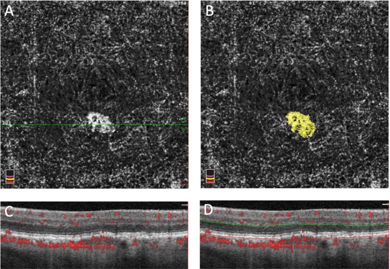
Purpose: To use optical coherence tomography angiography (OCTA) to study longitudinal subclinical choroidal neovascularization (CNV) changes and their correlation with progression to exudation in age-related macular degeneration (AMD).
Methods: This study included a total of 34 patients with unilateral neovascular AMD who were evaluated prospectively using OCTA to detect subclinical CNV in their fellow eye. Eyes with baseline subclinical CNV were followed with serial OCTA for a minimum of one year (15.2±3.27 months) to monitor the development of exudation.
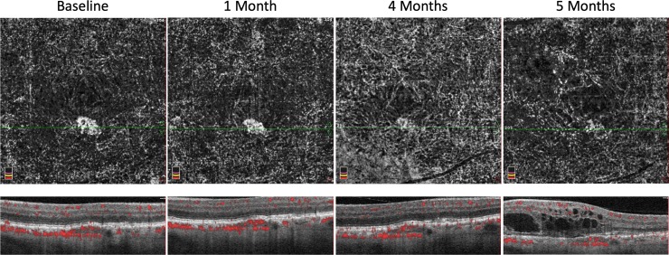
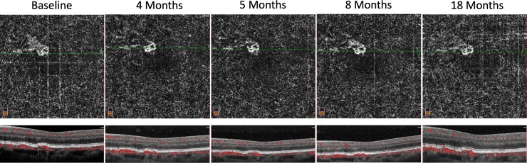
Results: Of the 34 fellow eyes studied, five were found to have baseline subclinical CNV. One of the five cases of baseline subclinical CNV converted to exudative AMD during the follow up period. The average surface area of baseline subclinical CNV on OCTA was 0.131±0.096 mm2 which progressed to 0.136±0.104 mm2 at the final follow up (P = 0.539). Geographic atrophy grew at a rate of 0.82±1.20mm2/year in four eyes without subclinical CNV and 0.02mm2/year in one eye with subclinical CNV.
Conclusion and importance: The rate of conversion to exudative AMD in eyes with subclinical CNV of 20% in our study is similar to previous reports and suggests the importance of vigilance in these eyes. The lower growth rate of geographic atrophy may suggest a protective effect of subclinical CNV that deserves further study.
Conflict of interest statement
The authors have declared that no competing interests exist.
Figures
References
Publication types
MeSH terms
Grants and funding
LinkOut - more resources
Full Text Sources
Medical
