Review
. 2019 Jun 7:16:791-804.
doi: 10.1016/j.omtn.2019.04.027. Epub 2019 May 15.

Emerging Function and Clinical Values of Exosomal MicroRNAs in Cancer

Affiliations
Review

Emerging Function and Clinical Values of Exosomal MicroRNAs in Cancer

Man Wang et al. Mol Ther Nucleic Acids. .

Abstract

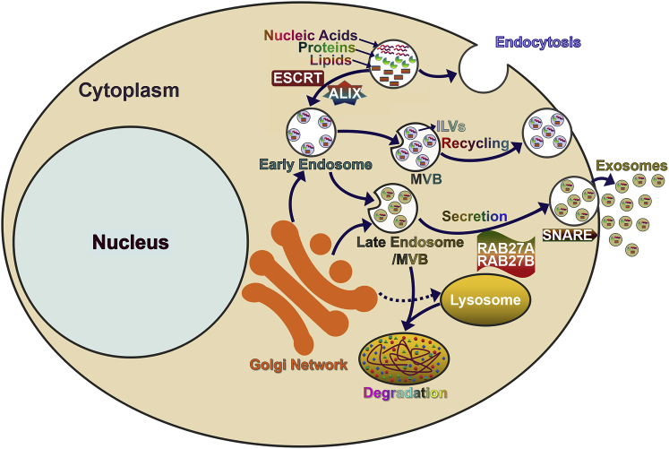
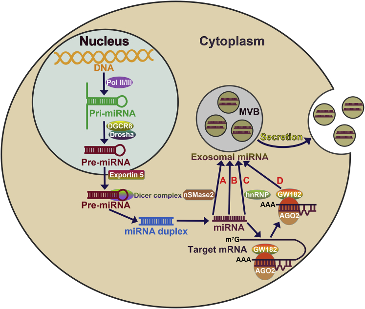
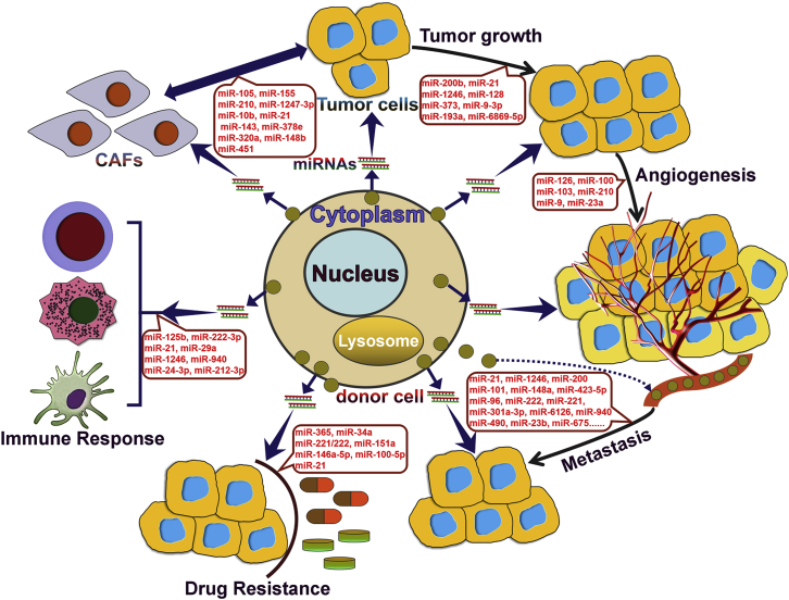
Exosomes are a subset of membrane-bound extracellular vesicles with diameters ranging from 30 to 100 nm. Exosomes enclose a variety of molecules, such as lipids, proteins, and non-coding RNAs. In the past decades, microRNAs (miRNAs) have attracted great attention in cancer research, as they play an important role in the occurrence and development of cancer. Increasing evidence indicates that tumor cells communicate with not only other tumor cells but also cells present in the tumor microenvironment via secretion and transfer of exosomal miRNAs. More importantly, exosomal miRNAs are found to serve as signaling molecules to regulate tumor growth, angiogenesis, metastasis, sensitivity to chemotherapy, and immune evasion. Deregulated expression of exosomal miRNAs is an early event in carcinogenesis and may reflect the malignant characteristics of cancer. Owing to the wide existence and high stability of exosomal miRNAs in body fluids, they may represent a novel class of non-invasive biomarkers for cancer. In this review, we highlight the recent advances on the functional role of exosomal miRNAs in cancer pathogenesis. We also discuss the potential clinical utility of exosome-shuttled miRNAs as biomarkers for the diagnosis and treatment of cancer.

Keywords: cancer biomarker; cancer diagnosis; cancer pathogenesis; cancer treatment; carcinogenesis; exosome; microRNA.


Figures

Figure 1
Figure 1
Schematic Representation of the Biogenesis and Secretion of Exosomes Exosomes are formed as intraluminal vesicles (ILVs). The cargoes (nucleic acids, proteins, and lipids) are ingested by the cells through the endocytotic pathway and are then transported to early endosomes. The maturation of early endosomes gives rise to multivesicular bodies (MVBs), late endosomes containing numerous ILVs. During the process of ILV generation, nucleic acids (miRNAs, DNAs, and RNAs), proteins (cytoplasmic proteins, tetraspanins, and membrane receptors), and lipids (ceramides and cholesterol) are incorporated into exosomes. Several molecules are involved in the biogenesis of ILVs, such as ESCRT and ALIX. MVBs can fuse with the cellular membranes to release exosomes into the extracellular space. Alternatively, MVBs can fuse with lysosomes, which results in the degradation of engulfed contents. MVB fusion with the cellular membrane is a fine-tuned multistep process involving MVB trafficking along microtubules, docking at the cellular membrane, and fusion to the cellular membrane. Several Rab GTPases (Rab27a and Rab27b) are implicated in the transport of MVBs to the cellular membrane and in the release of exosomes. Additionally, SNARE complexes may facilitate the fusion of MVBs with the cellular membranes. Exosomal cargoes can be delivered to the recipient cells via endocytosis, fusion with the cellular membrane, or ligand-receptor interaction.
Figure 2
Figure 2
Biogenesis, Packaging, and Secretion of Exosomal miRNAs miRNA genes are initially transcribed into primary miRNAs (pri-miRNAs) in the nucleus. A nuclear complex consisting of Drosha and DGCR8 cleaves the pri-miRNAs, resulting in the generation of precursor miRNAs (pre-miRNAs). Exportin 5 is responsible for nuclear export of the pre-miRNAs. In the cytoplasm, the pre-miRNAs are further processed by the Dicer complex into double-strand miRNAs (∼22 nt). One strand of the miRNA duplex (the mature miRNA) is selected to be incorporated into the RNA-induced silencing complex (RISC). The main components of the miRISC include miRNA, miRNA-targeted mRNA, GW182, and Argonaute 2 (Ago2). The miRNA in the miRISC can bind to the complementary sequence in the 3′ UTR of its target mRNA, which causes mRNA destabilization and translation suppression. Mature miRNAs are generally sorted into exosomes via four potential pathways: A, nSMase2-dependent pathway; B, the 3′ end of the miRNA sequence-dependent pathway (the 3′ end of the miRNA sequence contains an important sorting signal and thus guides specific miRNAs to be packed into exosomes); C, the miRNA motif and sumoylated hnRNP-dependent pathway (three hnRNP family proteins, hnRNPA2B1, hnRNPA1, and hnRNPC, guide miRNA loading into exosomes, and, among them, sumoylated hnRNPA2B1 specifically recognizes the GGAG motif in the 3′ portion of miRNA sequences and governs the sorting of miRNAs into exosomes); and D, the miRISC-related pathway. The RISC can co-localize with MVBs. Ago2 and miRNA-targeted mRNA are also associated with the exosomal assortment of miRNAs. Finally, MVBs fuse with the cellular membrane and release exosomes containing miRNAs into the extracellular space.
Figure 3
Figure 3
Summary of the Function of Exosomal miRNAs in Cancer Exosomal miRNAs play multifaceted roles in tumor initiation and development. Tumor cells could transfer exosomal miRNAs to surrounding tumor cells. In recipient tumor cells, exosomal miRNAs can control cell proliferation, invasion, and metastasis by orchestrating their downstream targets. Exosomal miRNAs mediate a special communication between tumor cells and endothelial cells, thus playing an important role in tumor angiogenesis. Drug-resistant tumor cells can transmit the resistant phenotype to drug-sensitive tumor cells through the horizontal transfer of exosomal miRNAs. In terms of mechanism, exosomal miRNAs mainly control the apoptotic or autophagic pathways, hence conferring chemoresistance to recipient tumor cells. Exosomal miRNAs serve as key messengers for exchanging information between tumor cells and immune cells (macrophages, T cells, and dendritic cells), contributing to the formation of a tumor-promoting, immunosuppressive microenvironment. In addition, exosomal miRNAs are instrumental for the cross-talk between tumor cells and CAFs within the tumor microenvironment. Tumor-derived exosomal miRNAs are capable of reprogramming and activating CAFs, thus facilitating tumorigenesis and tumor development. Exosomal miRNAs derived from adjacent CAFs in turn modulate the malignant phenotype of tumor cells.

Similar articles

Cited by

References

    1. Abak A., Abhari A., Rahimzadeh S. Exosomes in cancer: small vesicular transporters for cancer progression and metastasis, biomarkers in cancer therapeutics. PeerJ. 2018;6:e4763. - PMC - PubMed
    2. Abak, A., Abhari, A., and Rahimzadeh, S. (2018). Exosomes in cancer: small vesicular transporters for cancer progression and metastasis, biomarkers in cancer therapeutics. PeerJ 6, e4763. - PMC - PubMed
    1. Pap E., Pállinger E., Falus A. The role of membrane vesicles in tumorigenesis. Crit. Rev. Oncol. Hematol. 2011;79:213–223. - PubMed
    2. Pap, E., Pallinger, E., and Falus, A. (2011). The role of membrane vesicles in tumorigenesis. Crit. Rev. Oncol. Hematol. 79, 213-223. - PubMed
    1. van Niel G., Raposo G., Candalh C., Boussac M., Hershberg R., Cerf-Bensussan N., Heyman M. Intestinal epithelial cells secrete exosome-like vesicles. Gastroenterology. 2001;121:337–349. - PubMed
    2. van Niel, G., Raposo, G., Candalh, C., Boussac, M., Hershberg, R., Cerf-Bensussan, N., and Heyman, M. (2001). Intestinal epithelial cells secrete exosome-like vesicles. Gastroenterology 121, 337-349. - PubMed
    1. Blanchard N., Lankar D., Faure F., Regnault A., Dumont C., Raposo G., Hivroz C. TCR activation of human T cells induces the production of exosomes bearing the TCR/CD3/zeta complex. J. Immunol. 2002;168:3235–3241. - PubMed
    2. Blanchard, N., Lankar, D., Faure, F., Regnault, A., Dumont, C., Raposo, G., and Hivroz, C. (2002). TCR activation of human T cells induces the production of exosomes bearing the TCR/CD3/zeta complex. J. Immunol. 168, 3235-3241. - PubMed
    1. Kharaziha P., Ceder S., Li Q., Panaretakis T. Tumor cell-derived exosomes: a message in a bottle. Biochim. Biophys. Acta. 2012;1826:103–111. - PubMed
    2. Kharaziha, P., Ceder, S., Li, Q., and Panaretakis, T. (2012). Tumor cell-derived exosomes: a message in a bottle. Biochim. Biophys. Acta 1826, 103-111. - PubMed

LinkOut - more resources