. 2018;40(5-6):534-546.
doi: 10.1159/000499960. Epub 2019 Jun 4.

Ascending Lipopolysaccharide-Induced Intrauterine Inflammation in Near-Term Rabbits Leading to Newborn Neurobehavioral Deficits

Affiliations

Ascending Lipopolysaccharide-Induced Intrauterine Inflammation in Near-Term Rabbits Leading to Newborn Neurobehavioral Deficits

Zhongjie Shi et al. Dev Neurosci. 2018.

Abstract

Background: Chorioamnionitis from ascending bacterial infection through the endocervix is a potential risk factor for cerebral palsy. Tetrahydrobiopterin, an essential cofactor for nitric oxide synthase (NOS) and amino acid hydroxylases, when augmented in the fetal brain, prevents some of the cerebral palsy-like deficits in a rabbit hypoxia-ischemia model.

Objectives: To study the effect of lipopolysaccharide (LPS)-induced intrauterine inflammation in preterm gestation on motor deficits in the newborn, and whether biosynthesis of tetrahydrobiopterin or inflammatory mediators is affected in the fetal brain.

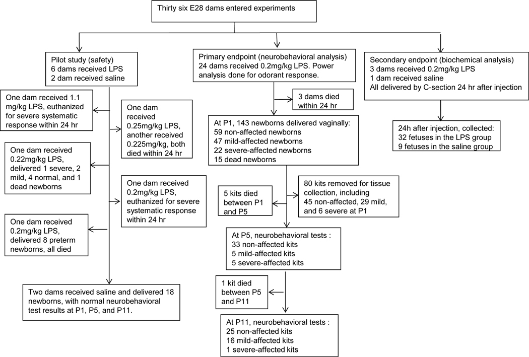
Methods: Pregnant rabbits at 28 days gestation (89% term) were administered either saline or LPS into both endocervical openings. One group underwent spontaneous delivery, and neurobehavioral tests were performed at postnatal day (P) 1 and P11, with some kits being sacrificed at P1 for histological analysis. Another group underwent Cesarean section 24 h after LPS administration. Gene sequences for rabbit biosynthetic enzymes of tetra-hydrobiopterin pathways were determined and analyzed in addition to cytokines, using quantitative real-time polymerase chain reaction.

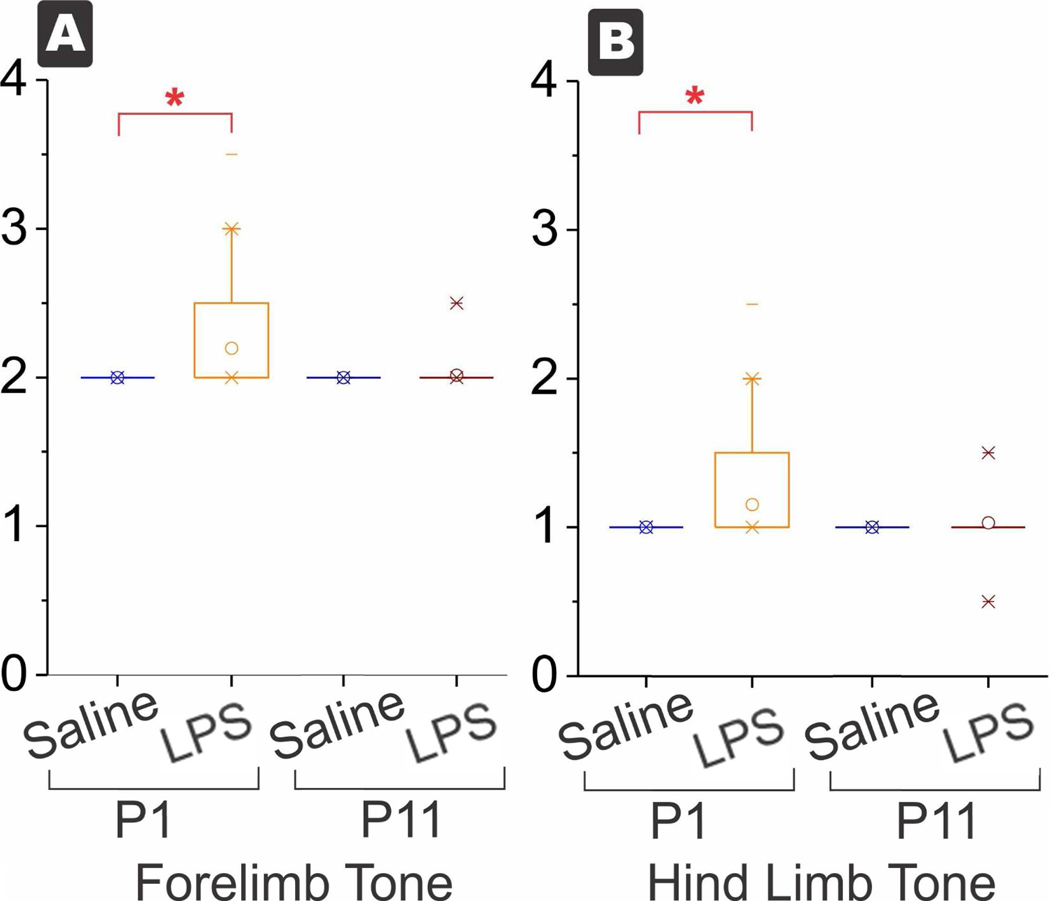
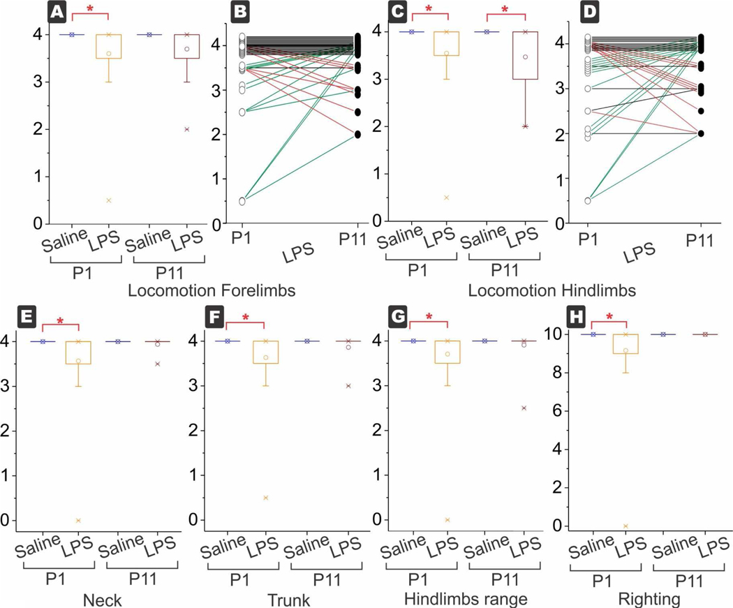
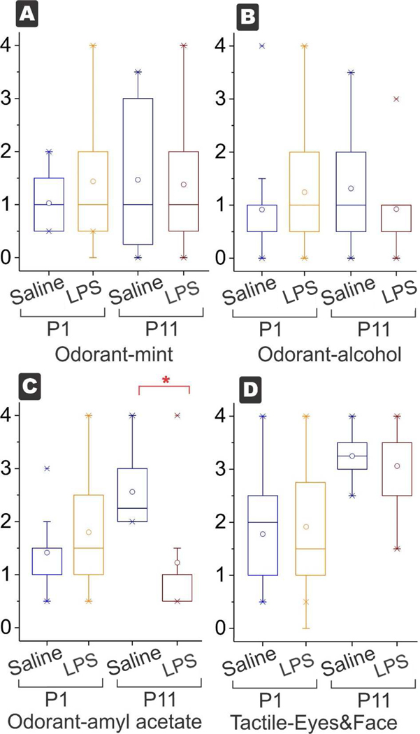
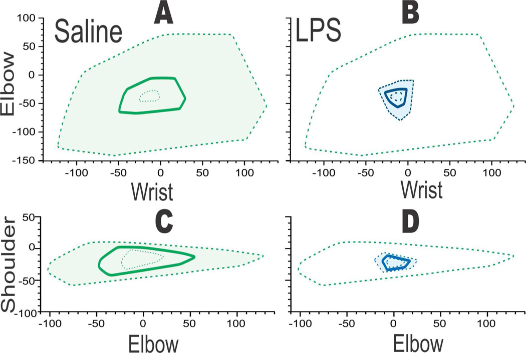
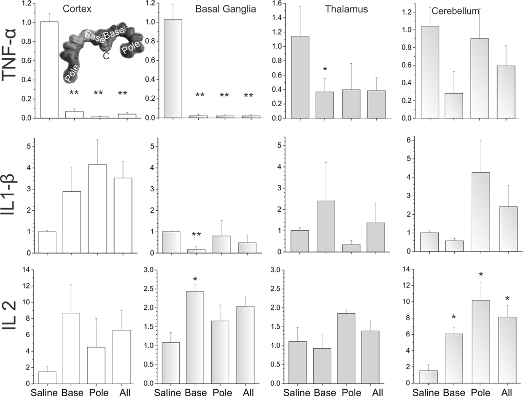
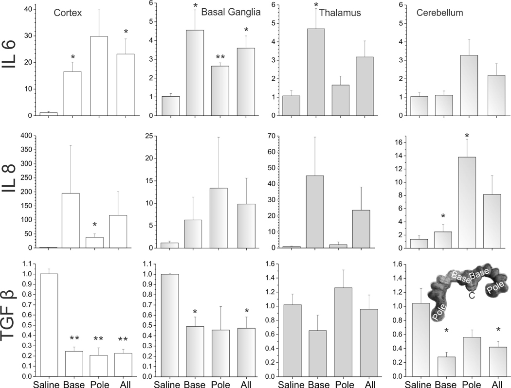
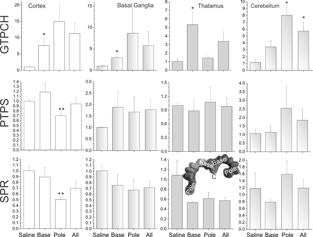
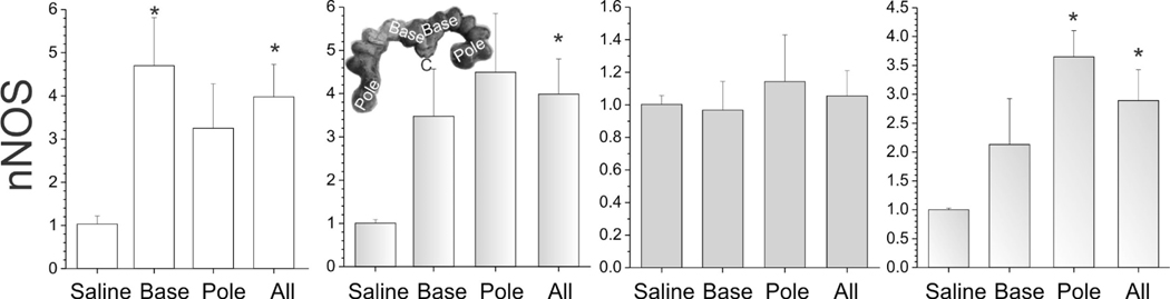
Results: Exposure to 200 μg/kg/mL LPS caused a locomotion deficit and mild hypertonia at P1. By P11, most animals turned into normal-appearing kits. There was no difference in neuronal cell death in the caudate between hypertonic and nonhypertonic kits at P1 (n = 3-5 in each group). Fetal brain GTP cyclohydrolase I was increased, whereas sepiapterin reductase and 6-pyruvoyltetrahydropterin synthase were decreased, 24 h after LPS administration. Neuronal NOS was also increased. Regardless of the position in the uterus or the brain region, expression of TNF-α and TGF-β was decreased, whereas that of IL-1β, IL-6, and IL-8 was increased (n = 3-4 in each group).

Conclusions: This is the first study using an ascending LPS-induced intrauterine inflammation model in rabbits, showing mostly transient hypertonia and mainly locomotor deficits in the kits. Not all proinflammatory cytokines are increased in the fetal brain following LPS administration. Changes in key tetrahydro-biopterin biosynthetic enzymes possibly indicate different effects of the inflammatory insult.

Keywords: Cerebral palsy; Cytokine; Lipopolysaccharide; Neurobehavioral assessment; Tetrahydrobiopterin.


Conflict of interest statement

Conflict of Interest

The authors declare no conflicts of interest.

Figures

Figure 1.
Figure 1.
Experimental flow chart showing breakdown of number and outcome of dams and fetuses/kits.
Figure 2.
Figure 2.
Box and whisker plots of tone. Increased tone in forelimbs (A) and hind limbs (B) at P1 in the LPS group but the survivors at P11 were not significantly different between saline and LPS. *p<0.0038, with Bonferroni correction for multiple tests.
Figure 3.
Figure 3.
Box and whisker plots of each variable. Decreased motor function in locomotion of forelimbs (A) and hind limbs (C) at P1 in the LPS group. The locomotion of hind limbs was persistently low at P11, the only motor test to be significant at P11. In the LPS group, change in each kit is shown for forelimb in (B) and hind limb in (D). Arrows and symbols have been offset a little artificially to show change for each kit. Red lines show worsening and green lines show improvement from P1 to P11. The number of worsening kits equaled roughly the number of improving kits both in forelimbs and hind limbs, while the biggest proportion was in the kits that were unchanged. Motor function of neck (E), trunk (F), range of hind limbs (G) and righting (H) were all decreased at P1 and recovered to normal at P11. The forelimb range was not shown for brevity but was also decreased at P1. *p<0.0038, with Bonferroni correction for multiple tests.
Figure 4.
Figure 4.
Box and whisker plots for sensory variables. Odorant response to peppermint (A) and alcohol (B) were not different at P1 or P11 (power=71% with moderate sized effect and α error=0.05). The only decrease was observed in odorant amyl acetate (C) at P11 but not at P1. (*p<0.0038, with Bonferroni correction for multiple tests). There was no difference between the tactile response to eye and face (D) at P1 and P11 (power=71% with moderate sized effect and α error=0.05).
Figure 5.
Figure 5.
Two joint analysis showing deficit in wrist-elbow and elbow-shoulder range of motion when saline (A,C green) was compared to LPS (B, D blue, n=3/group). Saline or LPS 0.2 mg/kg administered intracervically at E28 (89% gestation. After delivery, subtle locomotor deficits noticed at P1. Polygons showing mean (bold) with 95% confidence intervals in dashed lines. Green dashed line in B,D for reference of upper 95%tile of saline.
Figure 6.
Figure 6.
The expression of cytokines in different brain regions of E29 fetus. TNF-α, IL-1 β, IL-2. The unit of Y-axis is fold change, with n=3–4 in each group. The 4 columns represent the saline group, the base group, the pole group, and LPS group (all=base + pole), respectively. *p<0.05; **p<0.01, compared with the saline group.
Figure 7.
Figure 7.
The expression of cytokines in different brain regions of E29 fetus. IL-6, IL-8 and TGF-β. The unit of Y-axis is fold change, with n=3–4 in each group. The 4 columns represent the saline group, the base group, the pole group, and LPS group (all=base + pole), respectively. *p<0.05; **p<0.01, compared with the saline group.
Figure 8.
Figure 8.
The expression of BH4 biosynthesis enzymes in different brain regions of E29 fetus: GTPCH, PTPT, and SPR. The unit of Y-axis is fold change, with n=3–4 in each group. The 4 columns represent the saline group, the base group, the pole group, and LPS group (all=base + pole), respectively. *p<0.05; **p<0.01, compared with the saline group.
Figure 9.
Figure 9.
The expression of nNOS in different brain regions of E29 fetus. The unit of Y-axis is fold change, with n=3–4 in each group. The 4 columns represent the saline group, the base group, the pole group, and LPS group (all=base + pole), respectively. *p<0.05; **p<0.01, compared with the saline group.

References

    1. Graham HK, Rosenbaum P, Paneth N, Dan B, Lin JP, Damiano DL, Becher JG, Gaebler-Spira D, Colver A, Reddihough DS, Crompton KE, Lieber RL. Cerebral palsy. Nat Rev Dis Primers 2016;2:15082. - PMC - PubMed
    1. Koman LA, Smith BP, Shilt JS: Cerebral palsy. Lancet 2004;363:1619–1631. - PubMed
    1. Yoon BH, Park CW, Chaiworapongsa T: Intrauterine infection and the development of cerebral palsy. BJOG 2003;110 Suppl 20:124–127. - PubMed
    1. Saadani-Makki F, Kannan S, Lu X, Janisse J, Dawe E, Edwin S, Romero R, Chugani D : Intrauterine administration of endotoxin leads to motor deficits in a rabbit model: a link between prenatal infection and cerebral palsy. Am J Obstet Gynecol 2008;199:651.e1-e7. - PMC - PubMed
    1. Yu L, Vásquez-Vivar J, Jiang R, Luo K, Derrick M, Tan S: Developmental susceptibility of neurons to transient tetrahydrobiopterin insufficiency and antenatal hypoxia-ischemia in fetal rabbits. Free Radic Biol Med 2014;67:426–436. - PMC - PubMed