

Three Melanin Pathway Genes, TH, yellow, and aaNAT, Regulate Pigmentation in the Twin-Spotted Assassin Bug, Platymeris biguttatus (Linnaeus)
- PMID: 31163651
- PMCID: PMC6600426
- DOI: 10.3390/ijms20112728
Three Melanin Pathway Genes, TH, yellow, and aaNAT, Regulate Pigmentation in the Twin-Spotted Assassin Bug, Platymeris biguttatus (Linnaeus)
Abstract
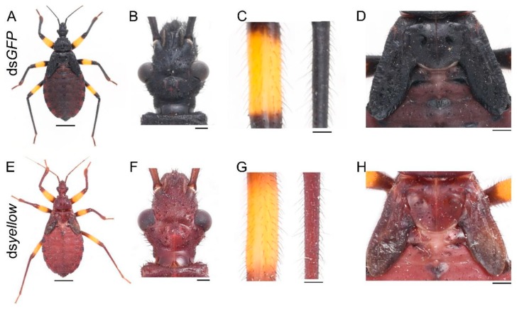
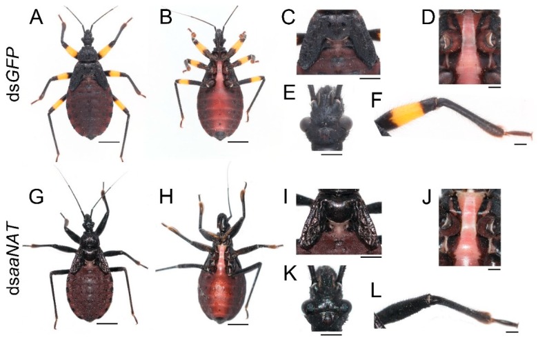
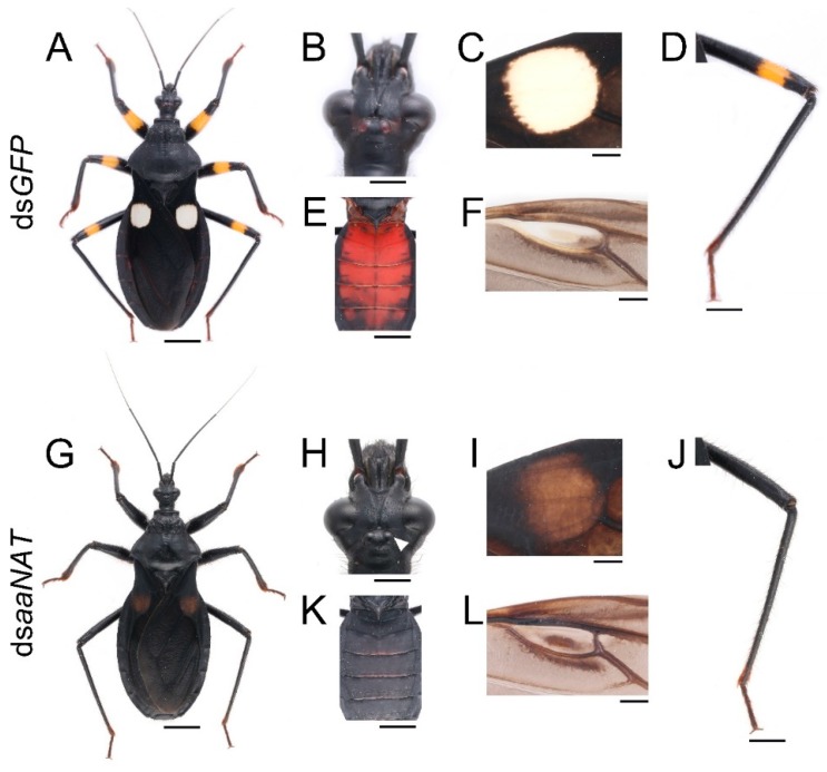
Pigmentation plays a vital role in insect survival and reproduction. Many melanin pathway genes have been studied in holometabolous insects; however, they have only been studied in two hemimetabolous insect genera, Oncopeltus and Periplaneta. Here we analyzed three melanin pathway genes (TH, yellow, and aaNAT) using RNA interference (RNAi) in another hemimetabolous insect, namely the twin-spotted assassin bug, Platymeris biguttatus. TH was highly expressed in freshly molted nymphs and adults. TH RNAi resulted in a complete loss of black pigment, with yellow coloration maintained. Therefore, black pigment in this assassin bug is solely generated from the melanin pathway, whereas yellow pigment is generated from other unknown pigmentation pathways. yellow and aaNAT were highly expressed in the white spot of the hemelytra. Downregulation of yellow caused a brown phenotype with high mortality, indicating an important role of yellow functions in cuticle formation and in the process of converting melanin from brown to black. Interestingly, aaNAT RNAi caused not only loss of white pigment, but also loss of yellow and red pigments. This phenotype of aaNAT has not been reported in other insects. Our results provide new information for understanding the melanin pathway in which aaNAT is essential for the formation of colorless patterns.
Keywords: Platymeris biguttatus; arylalkylamine-N-acetyltransferase (aaNAT); pigmentation; tyrosine hydroxylase (TH); yellow.
Conflict of interest statement
The authors declare no conflict of interest.
Figures


Similar articles
-
A Pathway Analysis of Melanin Patterning in a Hemimetabolous Insect.Genetics. 2016 May;203(1):403-13. doi: 10.1534/genetics.115.186684. Epub 2016 Mar 16. Genetics. 2016. PMID: 26984060 Free PMC article.
-
Disruption of an N-acetyltransferase gene in the silkworm reveals a novel role in pigmentation.Development. 2010 Dec;137(23):4083-90. doi: 10.1242/dev.053678. Development. 2010. PMID: 21062865
-
The genetic control of aposematic black pigmentation in hemimetabolous insects: insights from Oncopeltus fasciatus.Evol Dev. 2014 Sep;16(5):270-7. doi: 10.1111/ede.12090. Epub 2014 Aug 14. Evol Dev. 2014. PMID: 25124093 Free PMC article.
-
Enzymatic control of pigmentation in mammals.FASEB J. 1991 Nov;5(14):2902-9. FASEB J. 1991. PMID: 1752358 Review.
-
Pigmentation and behavior: potential association through pleiotropic genes in Drosophila.Genes Genet Syst. 2013;88(3):165-74. doi: 10.1266/ggs.88.165. Genes Genet Syst. 2013. PMID: 24025245 Review.
Cited by
-
Atypical strategies for cuticle pigmentation in the blood-feeding hemipteran Rhodnius prolixus.Genetics. 2022 May 31;221(2):iyac064. doi: 10.1093/genetics/iyac064. Genetics. 2022. PMID: 35445704 Free PMC article.
-
Evolutionary Genomics Reveals Multiple Functions of Arylalkylamine N-Acetyltransferase in Fish.Front Genet. 2022 May 19;13:820442. doi: 10.3389/fgene.2022.820442. eCollection 2022. Front Genet. 2022. PMID: 35664299 Free PMC article. Review.
-
Gene Editing and Genetic Control of Hemipteran Pests: Progress, Challenges and Perspectives.Front Bioeng Biotechnol. 2022 Jun 7;10:900785. doi: 10.3389/fbioe.2022.900785. eCollection 2022. Front Bioeng Biotechnol. 2022. PMID: 35747496 Free PMC article. Review.
-
Combinatorial expression of ebony and tan generates body color variation from nymph through adult stages in the cricket, Gryllus bimaculatus.PLoS One. 2023 May 18;18(5):e0285934. doi: 10.1371/journal.pone.0285934. eCollection 2023. PLoS One. 2023. PMID: 37200362 Free PMC article.
-
RNAi-Mediated Manipulation of Cuticle Coloration Genes in Lygus hesperus Knight (Hemiptera: Miridae).Insects. 2022 Oct 27;13(11):986. doi: 10.3390/insects13110986. Insects. 2022. PMID: 36354810 Free PMC article.
References
-
- True J.R. Insect melanism: The molecules matter. Trends Ecol. Evol. 2003;18:640–647. doi: 10.1016/j.tree.2003.09.006. - DOI
-
- Nijhout H.F. The Development and Evolution of Butterfly Wing Patterns. Smithsonian Institution Press; Washington, DC, USA: 1991. p. 297.
-
- Maranda B., Hodgetts R. A characterization of dopamine acetyltransferase in Drosophila melanogaster. Insect Biochem. 1977;7:33–43. doi: 10.1016/0020-1790(77)90054-3. - DOI
MeSH terms
Substances
Grants and funding
LinkOut - more resources
Full Text Sources
