. 2019 Aug 6;140(6):500-513.
doi: 10.1161/CIRCULATIONAHA.119.041059. Epub 2019 Jun 5.

HHIPL1, a Gene at the 14q32 Coronary Artery Disease Locus, Positively Regulates Hedgehog Signaling and Promotes Atherosclerosis

Affiliations

HHIPL1, a Gene at the 14q32 Coronary Artery Disease Locus, Positively Regulates Hedgehog Signaling and Promotes Atherosclerosis

Dimitra Aravani et al. Circulation. .

Abstract

Background: Genome-wide association studies have identified chromosome 14q32 as a locus for coronary artery disease. The disease-associated variants fall in a hitherto uncharacterized gene called HHIPL1 (hedgehog interacting protein-like 1), which encodes a sequence homolog of an antagonist of hedgehog signaling. The function of HHIPL1 and its role in atherosclerosis are unknown.

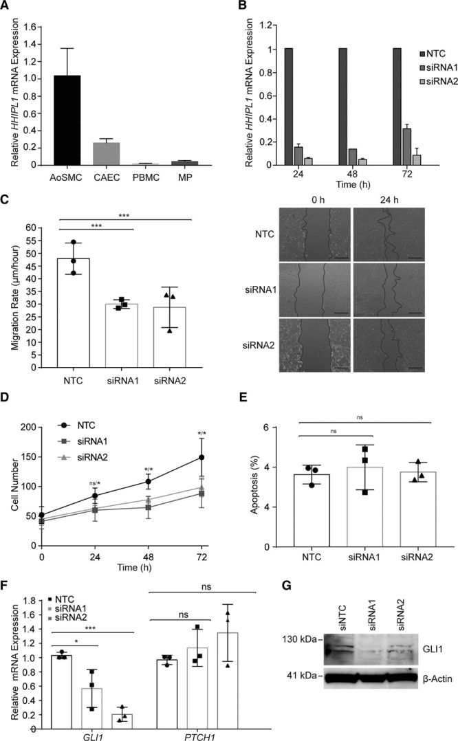
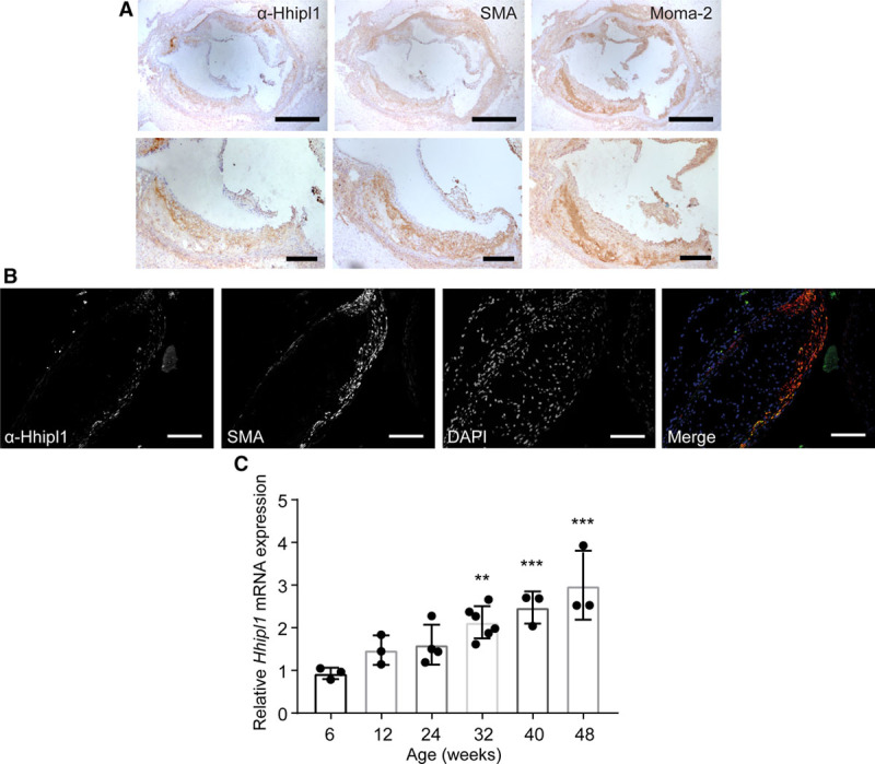
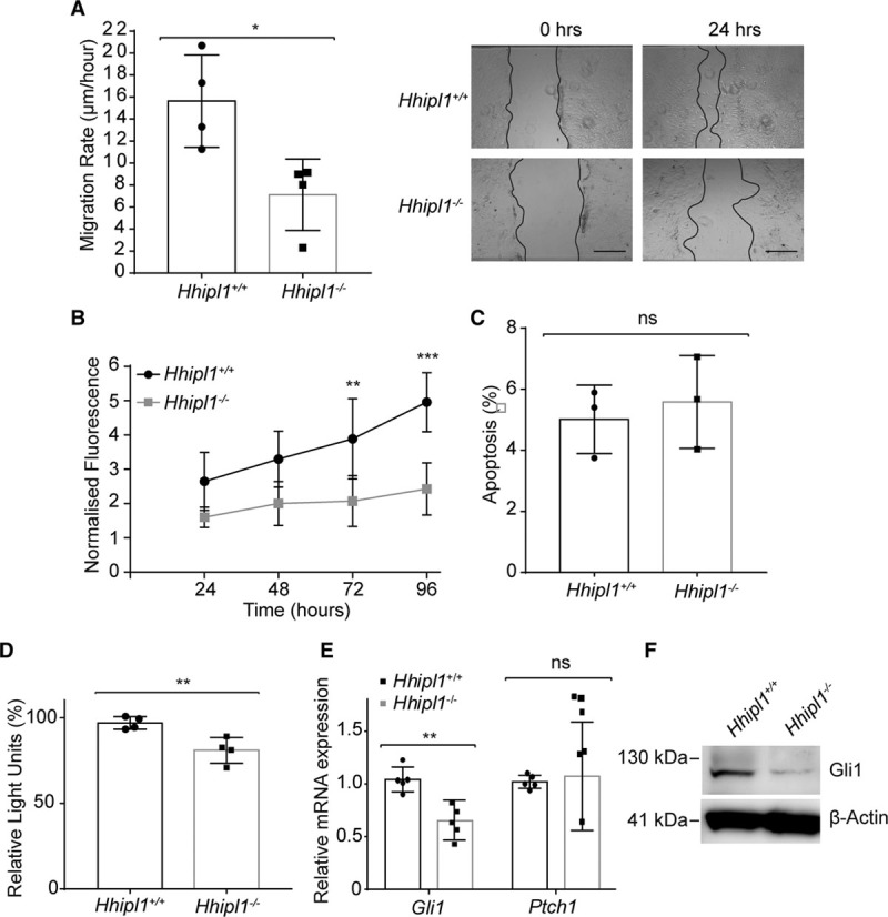
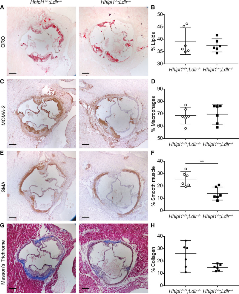
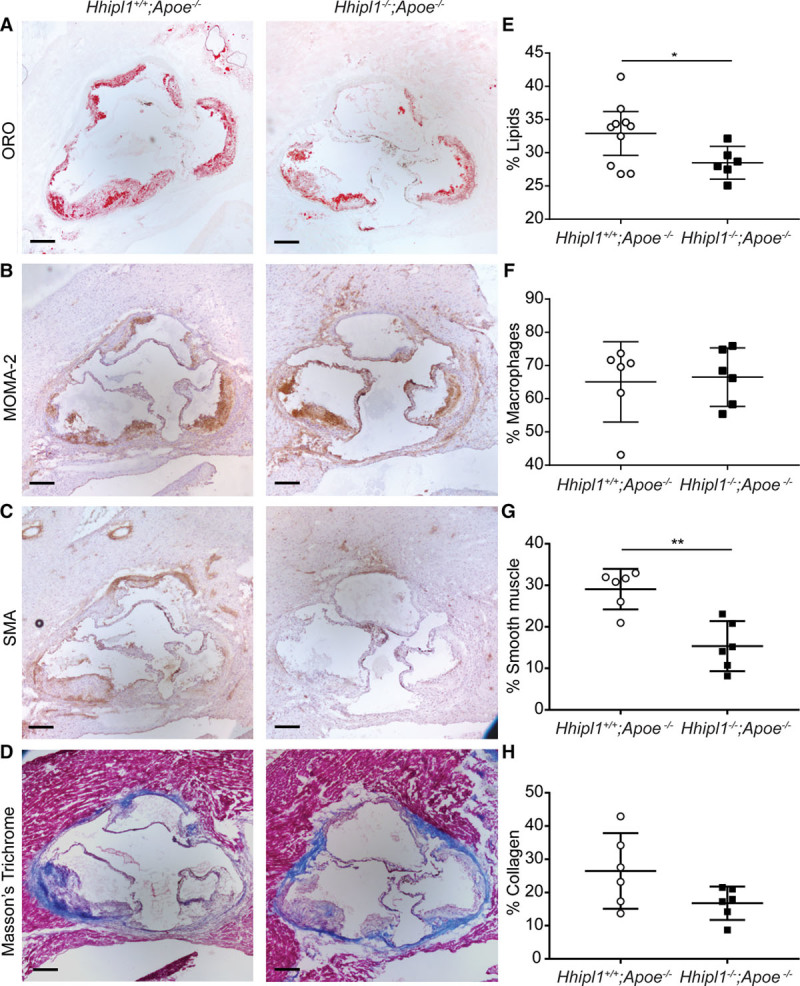
Methods: HHIPL1 cellular localization, interaction with sonic hedgehog (SHH), and influence on hedgehog signaling were tested. HHIPL1 expression was measured in coronary artery disease-relevant human cells, and protein localization was assessed in wild-type and Apoe-/- (apolipoprotein E deficient) mice. Human aortic smooth muscle cell phenotypes and hedgehog signaling were investigated after gene knockdown. Hhipl1-/- mice were generated and aortic smooth muscle cells collected for phenotypic analysis and assessment of hedgehog signaling activity. Hhipl1-/- mice were bred onto both the Apoe-/- and Ldlr-/- (low-density lipoprotein receptor deficient) knockout strains, and the extent of atherosclerosis was quantified after 12 weeks of high-fat diet. Cellular composition and collagen content of aortic plaques were assessed by immunohistochemistry.

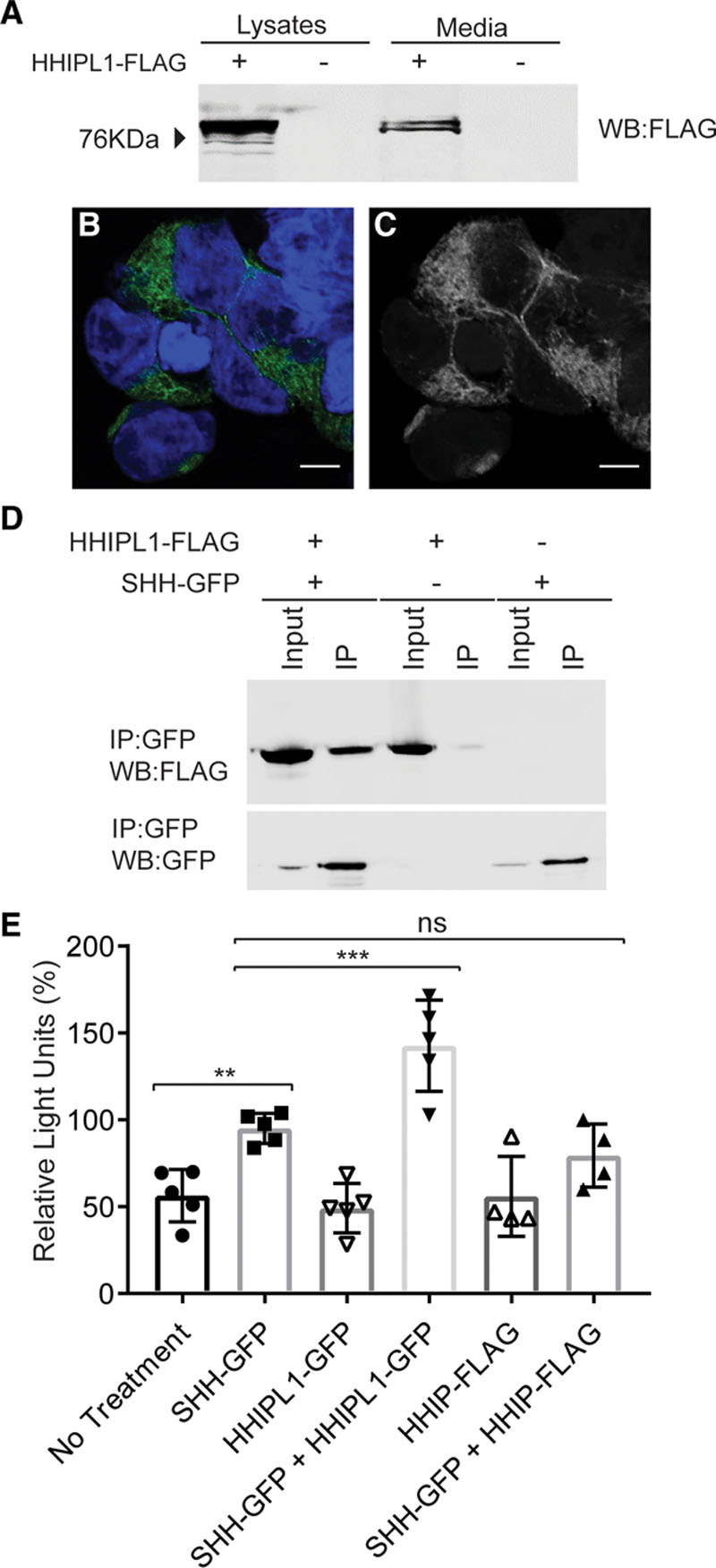
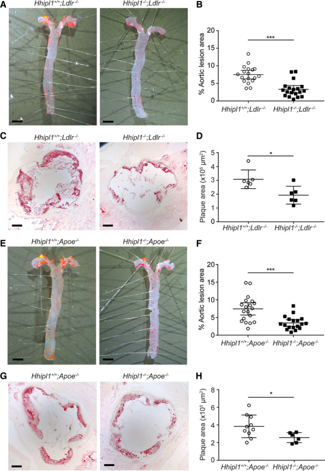
Results: In vitro analyses revealed that HHIPL1 is a secreted protein that interacts with SHH and increases hedgehog signaling activity. HHIPL1 expression was detected in human smooth muscle cells and in smooth muscle within atherosclerotic plaques of Apoe-/- mice. The expression of Hhipl1 increased with disease progression in aortic roots of Apoe-/- mice. Proliferation and migration were reduced in Hhipl1 knockout mouse and HHIPL1 knockdown aortic smooth muscle cells, and hedgehog signaling was decreased in HHIPL1-deficient cells. Hhipl1 knockout caused a reduction of >50% in atherosclerosis burden on both Apoe-/- and Ldlr-/- knockout backgrounds, and lesions were characterized by reduced smooth muscle cell content.

Conclusions: HHIPL1 is a secreted proatherogenic protein that enhances hedgehog signaling and regulates smooth muscle cell proliferation and migration. Inhibition of HHIPL1 protein function might offer a novel therapeutic strategy for coronary artery disease.

Keywords: atherosclerosis; coronary artery disease; genome-wide association study; hedgehogs; signaling.


Figures

Figure 1.
Figure 1.
HHIPL1 is a secreted interactor of SHH. A, Representative Western blot of HEK293 cell lysates and conditioned media after transfection of HHIPL1-FLAG plasmid. B and C, Representative confocal images of HEK293 cells expressing HHIPL1-GFP (green) costained with DAPI (blue). Bars, 10 μm. D, Western blots, immunoblotted with anti-GFP and anti-FLAG antibodies, after transfection of HEK293 cells with HHIPL1-FLAG, SHH-GFP, and immunoprecipitation with anti-GFP beads. E, Gli-luciferase activity in SHH-LIGHT2 cells incubated with conditioned media from HEK293 cells transfected with SHH-GFP, HHIPL1-GFP, or HHIP-FLAG or a mixture of SHH-GFP with HHIPL1-GFP or HHIPL-FLAG; n=4–5. GFP indicates green fluorescent protein; HHIPL1, hedgehog interacting protein-like 1; IP, immunoprecipitation; ns, nonsignificant; and SHH, sonic hedgehog. Error bars represent mean±SD. **P≤0.01, ***P≤0.001.
Figure 2.
Figure 2.
HHIPL1 regulates human AoSMC migration and proliferation. A,HHIPL1 mRNA expression in human aortic smooth muscle cells (AoSMC), coronary artery endothelial cells (CAEC), peripheral blood mononuclear cells (PBMC), and macrophages (MP) relative to 36B4. B,HHIPL1 mRNA expression relative to RPLP0 in AoSMCs 24 to 72 hours after transfection. C,Migration rate of rate of human AoSMCs after siRNA transfection (left; n=3). Representative images of wound-healing assay (right). D, Number of AoSMCs over 72 hours after siRNA knockdown. E,Proportion of apoptotic cells 48 hours after knockdown. F, GLI1 and PTCH1 expression relative to RPLP0 48 hours after siRNA knockdown. G, Representative Western blot of GLI1 after siRNA knockdown. β-actin was used as a loading control. AoSMC indicates aortic smooth muscle cell; HHIPL1, hedgehog interacting protein-like 1; NTC, nontargeting control siRNA; and siRNA, small interfering RNA.
Figure 3.
Figure 3.
Hhipl1 expression in atherosclerotic plaques. A, Representative immunohistochemical staining with anti–α smooth muscle actin antibody (SMA), anti-Hhipl1, and MOMA-2 in aortic root lesions from 18-week-old Apoe−/− mice fed Western diet for 12 weeks. Bars, 500 μm (top) and 200 μm (bottom). B, Immunofluorescent staining of aortic root lesion with DAPI, SMA, and anti-Hhipl1. Bars, 100 μm. C, Hhipl1 mRNA expression relative to Rpl4 in the aortic arch of 6- to 48-week-old Apoe−/− mice. n=3–6 mice per time point. Error bars represent mean±SD. HHIPL1 indicates hedgehog interacting protein-like 1. *Post hoc comparisons with 6-week time point. **P≤0.01, ***P≤0.001.
Figure 4.
Figure 4.
HHIPL1 regulates mouse AoSMC migration and proliferation. A,Migration rate of Hhipl1−/− and wild-type AoSMCs in a scratch wound assay over a period of 24 hours (n=4). Representative images are shown (right). B, Proliferation of Hhipl1−/− and wild-type AoSMCs over a period of 96 hours (n=4). *Significant post hoc comparisons at 72 and 96 hours. C, Proportion of apoptotic wild-type and Hhipl1−/− AoSMCs. D, Gli-luciferase activity in SHH-LIGHT2 cells co-cultured with either wild-type or Hhipl1−/− AoSMCs; n=4. E, Gli1 and Ptch1 mRNA expression relative to Rplp0 in wild-type and Hhipl1−/− AoSMCs; n=5. Error bars represent mean±SD. *P≤0.05, **P≤0.01, ***P≤0.001. F, Western blot showing Gli1 expression in wild-type and knockout cells. β-actin was used as a loading control. AoSMC indicates aortic smooth muscle cell; HHIPL1, hedgehog interacting protein-like 1; and SHH, sonic hedgehog.
Figure 5.
Figure 5.
Hhipl1 deficiency reduces atherosclerosis in Hhipl1−/−;Apoe−/− and Hhipl1−/−;Ldlr−/− mice. A, Representative Oil Red O–stained (ORO) aortas from Hhipl1+/+;Ldlr−/− and Hhipl1−/−;Ldlr−/− mice. B, Quantification of atherosclerosis in aortas of mice of each genotype as a percentage of total aorta area (n=18 vs n=19). C, Representative microphotographs of ORO-stained aortic root sections from Hhipl1+/+;Ldlr−/− and Hhipl1−/−;Ldlr−/− mice. D, Aortic root lesion area (9 sections per mouse, n=6 per group). E, ORO-stained aortas from Hhipl1+/+;Apoe−/− and Hhipl1−/−;Apoe−/− mice. F, Quantification of atherosclerosis in the aortas of mice of each genotype (n=19 vs n=18). G, ORO-stained aortic roots from Hhipl1+/+;Apoe−/− and Hhipl1−/−;Apoe−/− mice. H, Aortic root lesion area (9 sections per mouse, n=10 vs n=6). Bars, 2 mm (A and E); bars, 200 μm (C and G). Error bars represent mean±CI. HHIPL1 indicates hedgehog interacting protein-like 1. *P≤0.05, ***P≤0.001.
Figure 6.
Figure 6.
Hhipl1 deficiency reduces smooth muscle cell content in Hhipl1−/−;Ldlr−/−atherosclerotic lesions. Representative photomicrographs of atherosclerotic lesion components in Hhipl1+/+;Ldlr−/− and Hhipl1−/−;Ldlr−/− mice. A, Oil red O (ORO) staining for lipids; C, MOMA-2 staining for macrophages; E, anti–alpha smooth muscle actin (SMA) staining for smooth muscle cells; and (G) Masson trichrome for collagen. Bars, 200 μm. The percentage content (average of 9 sections per animal) of (B) lipids, (D) macrophages, (F) smooth muscle cells, and (H) collagen (n=6 per group). Error bars represent mean±CI. HHIPL1 indicates hedgehog interacting protein-like 1. **P≤0.01.
Figure 7.
Figure 7.
Hhipl1 deficiency reduces smooth muscle cell content in Hhipl1−/−;Apoe−/−atherosclerotic lesions. Representative photomicrographs of atherosclerotic lesion components in Hhipl1+/+;Apoe−/− and Hhipl1−/−;Apoe−/− mice. A, Oil red O (ORO) staining for lipids; C, MOMA-2 staining for macrophages; E, anti-α smooth muscle actin (SMA) staining for smooth muscle cells; and G, Masson’s trichrome for collagen. Bars, 200 μm. Percentage coverage (average of 9 sections per animal) of (B) lipids, (D) macrophages, (F) smooth muscle cells (P=0.001), and (H) collagen (n=6–10 per group). Error bars represent mean±CI. HHIPL1 indicates hedgehog interacting protein-like 1. *P≤0.05, **P≤0.01. Bar, 200 μm.

References

    1. Schunkert H, König IR, Kathiresan S, Reilly MP, Assimes TL, Holm H, Preuss M, Stewart AF, Barbalic M, Gieger C, et al. Cardiogenics; CARDIoGRAM Consortium. Large-scale association analysis identifies 13 new susceptibility loci for coronary artery disease. Nat Genet. 2011;43:333–338. doi: 10.1038/ng.784. - PMC - PubMed
    1. Nikpay M, Goel A, Won HH, Hall LM, Willenborg C, Kanoni S, Saleheen D, Kyriakou T, Nelson CP, Hopewell JC, et al. A comprehensive 1,000 Genomes-based genome-wide association meta-analysis of coronary artery disease. Nat Genet. 2015;47:1121–1130. doi: 10.1038/ng.3396. - PMC - PubMed
    1. Klarin D, Zhu QM, Emdin CA, Chaffin M, Horner S, McMillan BJ, Leed A, Weale ME, Spencer CCA, Aguet F, et al. CARDIoGRAMplusC4D Consortium. Genetic analysis in UK Biobank links insulin resistance and transendothelial migration pathways to coronary artery disease. Nat Genet. 2017;49:1392–1397. doi: 10.1038/ng.3914. - PMC - PubMed
    1. Nelson CP, Goel A, Butterworth AS, Kanoni S, Webb TR, Marouli E, Zeng L, Ntalla I, Lai FY, Hopewell JC, et al. EPIC-CVD Consortium; CARDIoGRAMplusC4D; UK Biobank CardioMetabolic Consortium CHD working group. Association analyses based on false discovery rate implicate new loci for coronary artery disease. Nat Genet. 2017;49:1385–1391. doi: 10.1038/ng.3913. - PubMed
    1. Katoh Y, Katoh M. Comparative genomics on HHIP family orthologs. Int J Mol Med. 2006;17:391–395. doi: 10.3892/ijmm.17.2.391. - PubMed

Publication types