

Methionine restriction delays senescence and suppresses the senescence-associated secretory phenotype in the kidney through endogenous hydrogen sulfide
- PMID: 31164038
- PMCID: PMC6619995
- DOI: 10.1080/15384101.2019.1618124
Methionine restriction delays senescence and suppresses the senescence-associated secretory phenotype in the kidney through endogenous hydrogen sulfide
Abstract
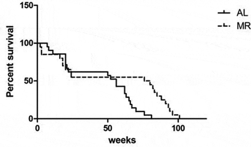
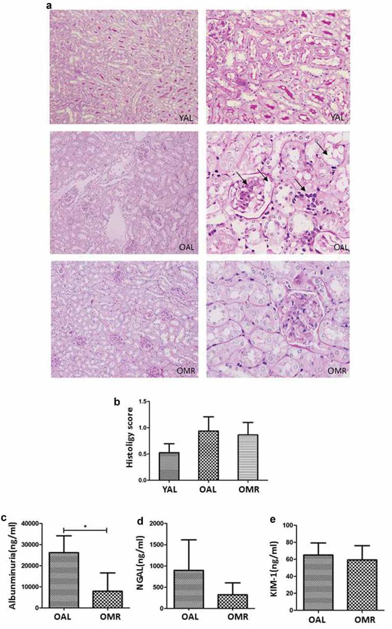
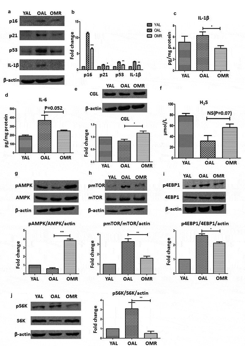
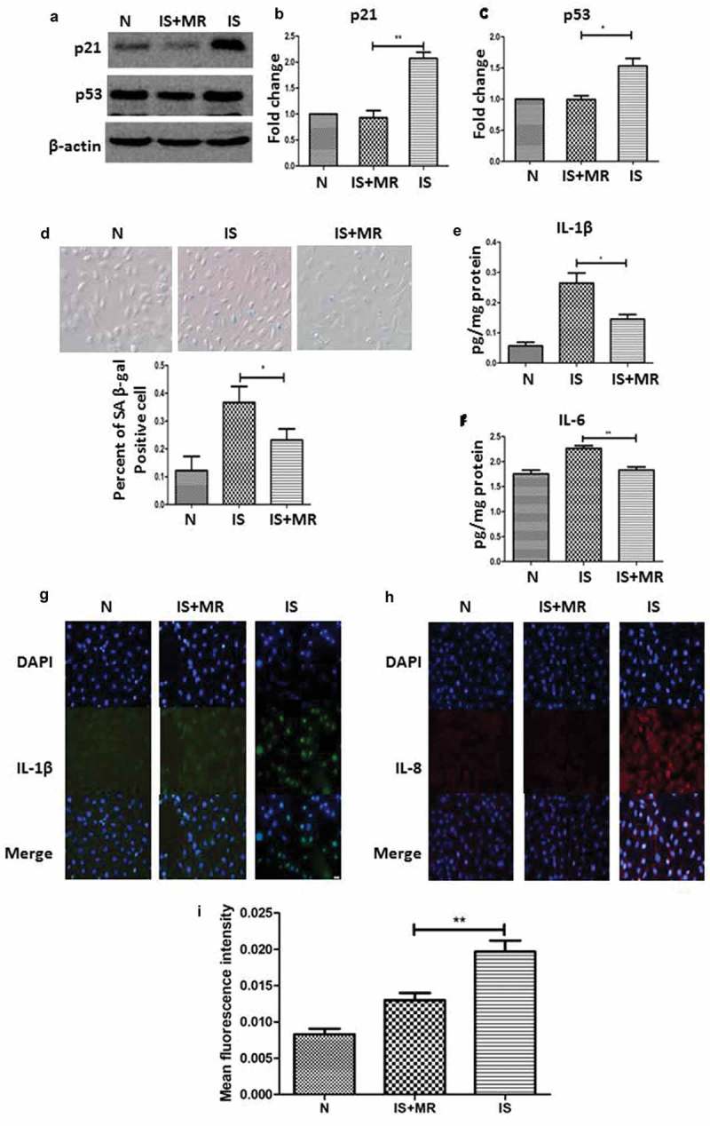
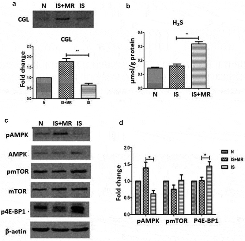
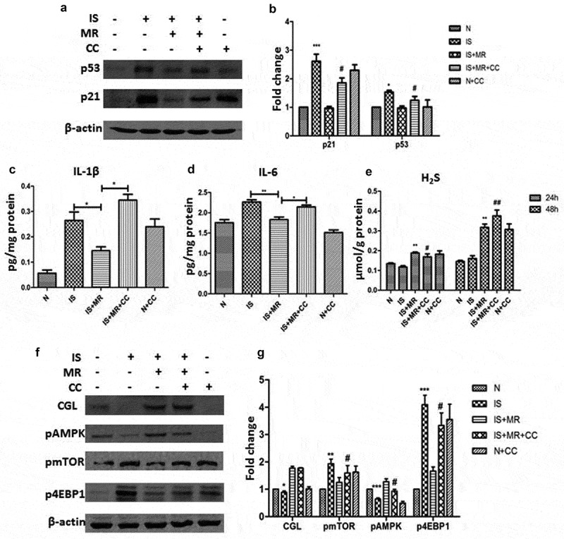
Aging is a risk factor for various acute and chronic kidney injuries. Kidney aging is accompanied by the secretion of growth factors, proteases, and inflammatory cytokines, known as the senescence-associated secretory phenotype (SASP). These factors accelerate the aging process and senescence-associated changes. Delaying kidney senescence may prevent acute and chronic kidney injury. Methionine restriction (MR) was found to be an effective intervention for delaying senescence. However, the mechanism of MR remains unclear. In this study, we investigated the effect of MR on the survival rate and renal aging of C57BL/6 mice and examined the relevant mechanisms. MR increased the survival rate and decreased the levels of senescence markers in the aging kidney. Both in vivo and in vitro, MR upregulated the transsulfuration pathway to increase H2S production, downregulated senescence markers and the SASP, and activated AMPK. The ability of MR to delay aging was reduced when AMPK was inhibited. These results suggest that MR may slow animal aging and kidney senescence through H2S production and AMPK pathway activation. Abbreviations: DR: diet restriction; MR: methionine restriction; SASP: senescence-associated secretory phenotype; AL: ad libitum; CKD, chronic kidney disease; AKI: acute kidney disease; TSP: transsulfuration pathway; CGL: cystathionine g-lyase; H2S: hydrogen sulfide; AMPK: AMP-activated protein kinase; mTOR: mammalian target of rapamycin; IS: indoxyl sulfate; CC: compound C.
Keywords: Senescence; hydrogen sulfide; methionine restriction; renal; senescence-associated secretory phenotype.
Figures
Similar articles
-
Hydrogen sulfide mediates the protection of dietary restriction against renal senescence in aged F344 rats.Sci Rep. 2016 Jul 26;6:30292. doi: 10.1038/srep30292. Sci Rep. 2016. PMID: 27456368 Free PMC article.
-
mTORC1-Sch9 regulates hydrogen sulfide production through the transsulfuration pathway.Aging (Albany NY). 2019 Oct 3;11(19):8418-8432. doi: 10.18632/aging.102327. Epub 2019 Oct 3. Aging (Albany NY). 2019. PMID: 31582588 Free PMC article.
-
Inhibition of endogenous hydrogen sulfide production reduces activation of hepatic stellate cells via the induction of cellular senescence.Cell Cycle. 2024 Mar;23(6):629-644. doi: 10.1080/15384101.2024.2345477. Epub 2024 Jun 5. Cell Cycle. 2024. PMID: 38836592 Free PMC article.
-
Calorie restriction and methionine restriction in control of endogenous hydrogen sulfide production by the transsulfuration pathway.Exp Gerontol. 2015 Aug;68:26-32. doi: 10.1016/j.exger.2014.12.010. Epub 2014 Dec 16. Exp Gerontol. 2015. PMID: 25523462 Free PMC article. Review.
-
Hydrogen sulfide in longevity and pathologies: Inconsistency is malodorous.Ageing Res Rev. 2021 May;67:101262. doi: 10.1016/j.arr.2021.101262. Epub 2021 Jan 28. Ageing Res Rev. 2021. PMID: 33516916 Review.
Cited by
-
The interaction between cellular senescence and chronic kidney disease as a therapeutic opportunity.Front Pharmacol. 2022 Aug 26;13:974361. doi: 10.3389/fphar.2022.974361. eCollection 2022. Front Pharmacol. 2022. PMID: 36091755 Free PMC article. Review.
-
Beneficial effects of time and energy restriction diets on the development of experimental acute kidney injury in Rat: Bax/Bcl-2 and histopathological evaluation.BMC Nephrol. 2023 Mar 20;24(1):59. doi: 10.1186/s12882-023-03104-6. BMC Nephrol. 2023. PMID: 36941590 Free PMC article.
-
Vitamin B Supplementation and Nutritional Intake of Methyl Donors in Patients with Chronic Kidney Disease: A Critical Review of the Impact on Epigenetic Machinery.Nutrients. 2020 Apr 27;12(5):1234. doi: 10.3390/nu12051234. Nutrients. 2020. PMID: 32349312 Free PMC article. Review.
-
Effect of Methionine Restriction on Aging: Its Relationship to Oxidative Stress.Biomedicines. 2021 Jan 29;9(2):130. doi: 10.3390/biomedicines9020130. Biomedicines. 2021. PMID: 33572965 Free PMC article. Review.
-
Cellular senescence of renal tubular epithelial cells in renal fibrosis.Front Endocrinol (Lausanne). 2023 Feb 28;14:1085605. doi: 10.3389/fendo.2023.1085605. eCollection 2023. Front Endocrinol (Lausanne). 2023. PMID: 36926022 Free PMC article. Review.
References
-
- Coresh J, Selvin E, Stevens LA, et al. Prevalence of chronic kidney disease in the United States. Jama. 2007;298:2038–2047. - PubMed
-
- Zhang L, Wang F, Wang L, et al. Prevalence of chronic kidney disease in China: a cross-sectional survey. Lancet. 2012;379:815–822. - PubMed
-
- Minutolo R, Borrelli S, De Nicola L.. CKD in the elderly: kidney senescence or blood pressure-related nephropathy? Am J Kidney Dis. 2015;66:184–186. - PubMed
-
- Coresh J, Astor BC, Greene T, et al. Prevalence of chronic kidney disease and decreased kidney function in the adult US population: third national health and nutrition examination survey. Am J Kidney Dis. 2003;41:1–12. - PubMed
Publication types
MeSH terms
Substances
LinkOut - more resources
Full Text Sources
Other Literature Sources
Medical
Miscellaneous