. 2019 Aug;9(8):1022-1035.
doi: 10.1158/2159-8290.CD-18-1494. Epub 2019 Jun 4.

Unique Neoantigens Arise from Somatic Mutations in Patients with Gastrointestinal Cancers

Affiliations

Unique Neoantigens Arise from Somatic Mutations in Patients with Gastrointestinal Cancers

Maria R Parkhurst et al. Cancer Discov. 2019 Aug.

Abstract

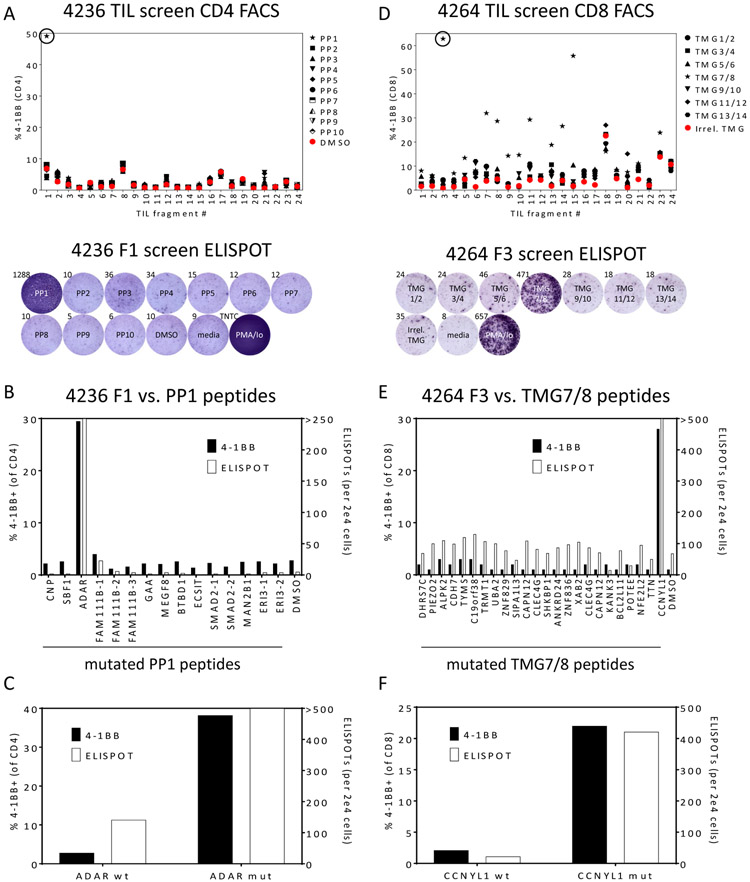
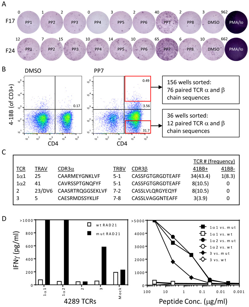
Immunotherapies can mediate regression of human tumors with high mutation rates, but responses are rarely observed in patients with common epithelial cancers. This raises the question of whether patients with these common cancers harbor T lymphocytes that recognize mutant proteins expressed by autologous tumors that may represent ideal targets for immunotherapy. Using high-throughput immunologic screening of mutant gene products identified via whole-exome sequencing, we identified neoantigen-reactive tumor-infiltrating lymphocytes (TIL) from 62 of 75 (83%) patients with common gastrointestinal cancers. In total, 124 neoantigen-reactive TIL populations were identified, and all but one of the neoantigenic determinants were unique. The results of in vitro T-cell recognition assays demonstrated that 1.6% of the gene products encoded by somatic nonsynonymous mutations were immunogenic. These findings demonstrate that the majority of common epithelial cancers elicit immune recognition and open possibilities for cell-based immunotherapies for patients bearing these cancers. SIGNIFICANCE: TILs cultured from 62 of 75 (83%) patients with gastrointestinal cancers recognized neoantigens encoded by 1.6% of somatic mutations expressed by autologous tumor cells, and 99% of the neoantigenic determinants appeared to be unique and not shared between patients.This article is highlighted in the In This Issue feature, p. 983.


Conflict of interest statement

Conflict of interest disclosure statement

The authors have no conflicts of interest to disclose.

Figures

Figure 1:
Figure 1:. Identification of neoantigen reactive CD4+ T cells from a patient with colon cancer (4236) and CD8+ T cells from a patient with esophageal cancer (4264).
(A) 24 TIL subcultures from patient 4236 were co-cultured overnight with autologous DCs pulsed with DMSO or the indicated peptide pool (PP) containing 15 or 16 peptides of 25 amino acids in length containing mutations identified by whole exome sequencing. TIL were also cultured without DCs (media) as negative controls or with PMA/Ionomycin (PMA/Io) as positive controls. T cell responses were measured by flow cytometric analysis of 4-1BB on CD4+ T cells (upper panel) and IFNγ ELISPOT (lower panel – one example showing the reactivity of the circled fragment in the upper panel). (B) TIL fragment culture F1 from patient 4236 was co-cultured overnight with autologous DCs pulsed with individual peptides from PP1, and T cell responses were measured by flow cytometric analysis of 4-1BB on CD4+ T cells (left axis) and IFNγ ELISPOT (right axis). By both criteria, F1 contained CD4+ T cells that recognized mutant ADAR. (C) 4236 TIL were co-cultured with autologous DCs pulsed with the indicated wild type and mutant peptides, and T cell responses were measured as in (B). (D) 24 TIL subcultures from patient 4264 were co-cultured overnight with autologous DCs transfected with an irrelevant TMG or the indicated TMG encoding mutations identified by whole exome sequencing. TIL were also cultured without DCs (media) as negative controls or with PMA/Ionomycin (PMA/Io) as positive controls. T cell responses were measured by flow cytometric analysis of 4-1BB on CD8+ T cells (upper panel) and IFNγ ELISPOT (lower panel – one example showing the reactivity of the circled fragment in the upper panel). (E) TIL fragment culture F3 from patient 4264 was co-cultured overnight with autologous DCs pulsed with individual 25 amino acid peptides derived from TMGs 7 and 8, and T cell responses were measured by flow cytometric analysis of 4-1BB on CD8+ T cells (left axis) and IFNγ ELISPOT (right axis). By both criteria, F3 contained CD8+ T cells that recognized mutant CCNYL1. (F) 4264 TIL were co-cultured with autologous DCs pulsed with the indicated wild type and mutant peptides, and T cell responses were measured as in (E).
Figure 2:
Figure 2:. Identification of neoantigen reactive TCRs from a patient with colon cancer (4289).
(A) 24 TIL subcultures from patient 4289 were co-cultured overnight with autologous DCs pulsed with DMSO or the indicated peptide pool (PP) containing 12 or 13 peptides of 25 amino acids in length containing mutations identified by whole exome sequencing. T cell responses were measured by flow cytometric analysis of 4-1BB on CD4+ T cells and IFNγ ELISPOT. ELISPOT results for 2 positive fragment cultures (F17 and F24) are shown. (B) T cells from F17, F24, and F18 (which recognized PP7 by flow cytometric analysis but not ELISPOT) were combined and co-cultured with autologous DCs pulsed with PP7. After gating on CD3+ cells, 156 individual CD4+ T cells that expressed high levels of 4-1BB, as indicated by the red box in the upper right hand panel of the FACS plot, were sorted into 96 well plate wells, and single cell RT-PCR was performed on the contents of each well to amplify TCR α and β chains. In addition, as negative controls, 36 individual CD4+ T cells that did not express 4-1BB, as indicated by the red box in the lower right hand panel of the FACS plot, were sorted into 96 well plate wells, and single cell RT-PCR was performed on the contents of each well to amplify TCR α and β chains. (C) Abbreviated sequences of the 5 most frequent TCRs identified by analysis of single-cell RT-PCR from CD4+ 4-1BB+ sorted cells in comparison to the frequencies of those TCRs in the CD4+ 4-1BB- population. (D) Autologous open-repertoire PBL were genetically modified via retroviral transduction to express the TCRs described in (C) and were then co-cultured overnight with DCs pulsed with all the wild type and mutant peptides from PP7. T cell responses were measured by IFNγ ELISA, and recognition of wild type and mutant RAD21 by T cells expressing each of the 5 TCRs is shown (left panel). Recognition of titrated amounts of wild type and mutant RAD21 25mers by T cells expressing the 3 reactive TCRs was evaluated by IFNγ ELISA (right panel).

References

    1. Monach PA, Meredith SC, Siegel CT, Schreiber H. A unique tumor antigen produced by a single amino acid substitution. Immunity 1995;2(1):45–59. - PubMed
    1. Matsushita H, Vesely MD, Koboldt DC, Rickert CG, Uppaluri R, Magrini VJ, et al. Cancer exome analysis reveals a T-cell-dependent mechanism of cancer immunoediting. Nature 2012;482(7385):400–4. - PMC - PubMed
    1. Lennerz V, Fatho M, Gentilini C, Frye RA, Lifke A, Ferel D, et al. The response of autologous T cells to a human melanoma is dominated by mutated neoantigens. Proc Natl Acad Sci U S A 2005;102(44):16013–8. - PMC - PubMed
    1. Hellmann MD, Nathanson T, Rizvi H, Creelan BC, Sanchez-Vega F, Ahuja A, et al. Genomic Features of Response to Combination Immunotherapy in Patients with Advanced Non-Small-Cell Lung Cancer. Cancer Cell 2018. May 14;33(5):843–852. - PMC - PubMed
    1. McGranahan N, Furness AJ, Rosenthal R, Ramskov S, Lyngaa R, Saini SK, et al. Clonal neoantigens elicit T cell immunoreactivity and sensitivity to immune checkpoint blockade. Science 2016;351(6280):1463–9. - PMC - PubMed

MeSH terms