. 2019 Aug 15;25(16):5135-5142.
doi: 10.1158/1078-0432.CCR-19-0271. Epub 2019 Jun 4.

Genomic Evolutionary Patterns of Leiomyosarcoma and Liposarcoma

Affiliations

Genomic Evolutionary Patterns of Leiomyosarcoma and Liposarcoma

Ali Amin-Mansour et al. Clin Cancer Res. .

Abstract

Purpose: Leiomyosarcoma and liposarcoma are common subtypes of soft tissue sarcoma (STS). Patients with metastatic leiomyosarcoma or dedifferentiated liposarcoma (DDLPS) typically have worse outcomes compared with localized leiomyosarcoma or well-differentiated liposarcoma (WDLPS). A better understanding of genetic changes between primary/metastatic leiomyosarcoma and between WDLPS/DDLPS may provide insight into their genetic evolution.

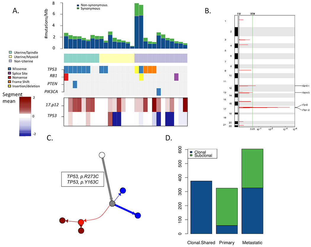
Experimental design: We interrogated whole-exome sequencing (WES) from "trios" of normal tissue, primary tumor, and metastatic tumor from individual patients with leiomyosarcoma (n = 9), and trios of normal tissue, well-differentiated tumor, and dedifferentiated tumor from individual patients with liposarcoma (n = 19). Specifically, we performed mutational, copy number, and tumor evolution analyses on these cohorts and compared patterns among leiomyosarcoma and liposarcoma trios.

Results: Leiomyosarcoma cases harbored shared drivers through a typical parent/child relationship where the metastatic tumor was derived from the primary tumor. In contrast, while all liposarcoma cases shared the characteristic focal chromosome 12 amplicon, most paired liposarcoma cases did not share additional mutations, suggesting a divergent evolutionary pattern from a common precursor. No highly recurrent genomic alterations from WES were identified that could be implicated as driving the progression of disease in either sarcoma subtype.

Conclusions: From a genomic perspective, leiomyosarcoma metastases contain genetic alterations that are also found in primary tumors. WDLPS and DDLPS, however, appear to divergently evolve from a common precursor harboring 12q amplification, rather than as a transformation to a higher-grade tumor. Further efforts to identify specific drivers of these distinct evolutionary patterns may inform future translational and clinical research in STS.


Figures

Figure 1:
Figure 1:. Genomic overview of matched primary (localized) and metastatic leiomyosarcomas.
(A) Overview of mutations observed in the 28 primary localized LMS samples, each from a unique patient. Each column represents a sample/patient. Mutation rates in the tumor samples are shown in the top barplot with reference to synonymous and non-synonymous mutation rates. LMS cases were classified into three subtypes of Uterine-Myxoid, Uterine-Spindle cell, and non-Uterine. Mutations in established biologically relevant genes in LMS (TP53, RB1, PTEN, PIK3CA, and BRAF) are shown. TP53 mutations were the most frequent somatic alterations observed. Similarly, the bottom track shows amplifications in chromosome 17p12. (B) Copy number alteration significance in primary LMS samples. Copy number alteration significance through GISTIC (24) in primary LMS samples demonstrate chromosome 17p12 is the only region that is significantly amplified (FDR<0.0001). (C) Phylogenetic tree of the primary and metastatic sample of patient 4. The white circle represents the germline sample, the grey sample shows the inferred parent clone of the tumor samples before there was divergence, the red circles represent the metastatic (sub)clones and the blue circles represent the primary (sub)clones. The length of the branches shows the proportion of mutations accumulated between each clone. (D) Barplot of the frequency of mutations demonstrates how many of the mutations were categorized as shared between primary and metastatic (Shared), exclusive to the primary samples or exclusive to the metastatic samples. The mutations have been also distinguished based on clonality. A considerable percentage of clonal somatic alterations is shared between the primary and metastatic samples.
Figure 2:
Figure 2:. Patient-matched WDLPS and DDLPS showing a distinct genomic pattern of tumor evolution.
(A) Gross resection specimen of a representative LPS sample highlights WD and DD components of a mixed liposarcoma. Histology images of WDLPS (B) and DDLPS (C) from patient 11 are shown. (D) Overlap of copy number changes in WD and DDLPS in chromosome 12 of patient 11 demonstrate high concordance between segmentations at those loci. Breakpoints occur in the same locations in both samples. CDK4 and MDM2 are highlighted. (E) Phylogenetic tree of the WD and DDLPS sample of patient 11. The white circle represents the germline sample, the grey sample shows the inferred parent clone of the tumor samples before there was divergence, the red circles represent the DD (sub)clones and the blue circles represent the WD (sub)clones. The length of the branches shows the proportion of mutations accumulated between each clone. (F) Rearrangements shared between the WD and DDLPS samples in patient 11. (G) Rearrangements unique to WDLPS. (H) Rearrangements unique to DDLPS.
Figure 3:
Figure 3:. Genomic overview of the 19 matched WD and DD liposarcoma samples.
(A) An overview of mutations in the 19 WD and DDLPS samples highlight mutation rate (top), established biologically relevant genes in LPS (middle) and focal amplifications involving MDM2/CDK4 (bottom). GISTIC copy number analysis demonstrates focal amplification in chromosome 12 in WD (B) and DD (C) LPS samples. (D) Overview of the frequencies of subclonal and clonal mutations that are either shared between primary and metastatic samples from the same individual or that are exclusive to either cohort. (E) Rare (~1%) point mutations are shared by the WD and DDLPS samples from the same patient.

References

    1. Barretina J, Taylor BS, Shantanu B, Ramos AH, Lagos-Quintana M, DeCarolis PL, et al. Subtype-specific genomic alterations define new targets for soft-tissue sarcoma therapy. Nature Genetics 42, 715–721 (2010). - PMC - PubMed
    1. Serrano C, George S Leiomyosarcoma In: Wagner AJ, editor. Hematology/Oncology Clinics of North America: Sarcoma. Philadelphia: Elsevier, Inc; 2013. p. 957–974. - PubMed
    1. Henze J, Bauer S, Liposarcomas In: Wagner AJ, editor. Hematology/Oncology Clinics of North America: Sarcoma. Philadelphia: Elsevier, Inc; 2013. p. 939–955. - PubMed
    1. Taylor BS, DeCarolis PL, Angeles CV, Brenet F, Schultz N, Antonescu CR, et al. Frequent alterations and epigenetic silencing of differentiation pathway genes in structurally rearranged liposarcomas. Cancer Discovery 1, 587–597 (2011). - PMC - PubMed
    1. Chibon F, Mariani O, Derré J, Mairal A, Coindre JM, Guillou L et al. ASK1 (MAP3K5) as a potential therapeutic target in malignant fibrous histiocytomas with 12q14-q15 and 6q23 amplifications. Genes, Chromosomes & Cancer 40, 32–37 (2004). - PubMed

Publication types