. 2019 Jun 4;9(1):8278.
doi: 10.1038/s41598-019-44569-6.

Surface modification of gold nanoparticles with neuron-targeted exosome for enhanced blood-brain barrier penetration

Affiliations

Surface modification of gold nanoparticles with neuron-targeted exosome for enhanced blood-brain barrier penetration

Mattaka Khongkow et al. Sci Rep. .

Abstract

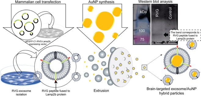
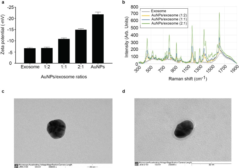
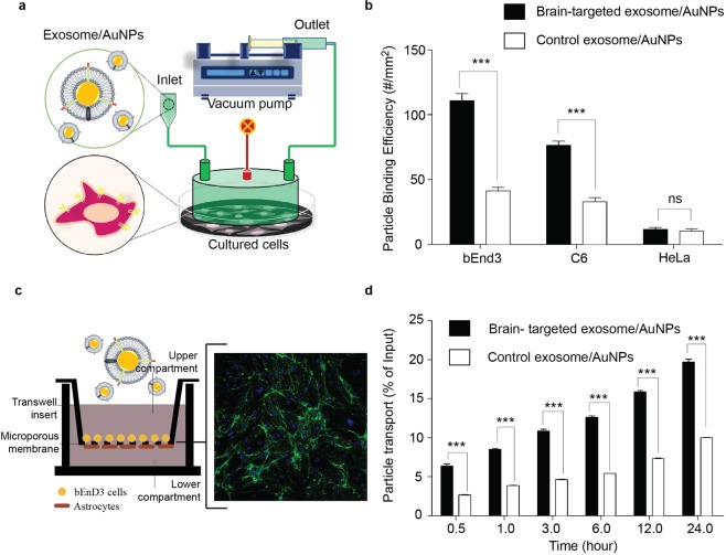
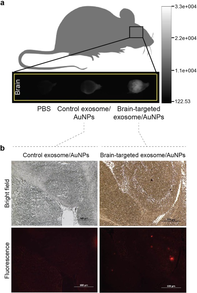
Gold nanoparticles (AuNPs) have been extensively used as nanomaterials for theranostic applications due to their multifunctional characteristics in therapeutics, imaging, and surface modification. In this study, the unique functionalities of exosome-derived membranes were combined with synthetic AuNPs for targeted delivery to brain cells. Here, we report the surface modification of AuNPs with brain-targeted exosomes derived from genetically engineered mammalian cells by using the mechanical method or extrusion to create these novel nanomaterials. The unique targeting properties of the AuNPs after fabrication with the brain-targeted exosomes was demonstrated by their binding to brain cells under laminar flow conditions as well as their enhanced transport across the blood brain barrier. In a further demonstration of their ability to target brain cells, in vivo bioluminescence imaging revealed that targeted-exosome coated AuNPs accumulated in the mouse brain after intravenous injection. The surface modification of synthetic AuNPs with the brain-targeted exosome demonstrated in this work represents a highly novel and effective strategy to provide efficient brain targeting and shows promise for the future in using modified AuNPs to penetrate the brain.


Conflict of interest statement

The authors declare no competing interests.

Figures

Figure 1
Figure 1
Schematic representation of gold nanoparticle surface functionalization through exosome coating. In this approach, exosome-producing cells were transiently transfected with a pcDNA GNSTM-3-RVG-10-Lamp2b-HA vector to express Lamp2b fusion protein fused in frame with the neuron-specific rabies viral glycoprotein (RVG) and glycosylation-stabilized (GNSTM) peptides, allowing the engineered cells to produce and secret exosomes with the RVG and GNSTM peptides on their surface as previously described by Hung, M. E., & Leonard, J. N., 2015. These engineered exosomes were collected then coated onto the surfaces of synthetic nanoparticles using the mechanical method or extrusion. Western blot of exosomes produced from transfected or non-transfected cells and probed with anti-HA antibody against HA tag present in Lamp2b fusion protein are also shown in the figure.
Figure 2
Figure 2
Physicochemical characteristics of the exosome-coated AuNPs. (a) The charge characteristics of exosome-coated AuNPs at different exosome/AuNPs ratios. (b) Raman spectra of exosome-coated AuNPs at different exosome/AuNPs ratios. (c) A representative TEM image of AuNP. (d) A representative TEM image of exosome-coated AuNP.
Figure 3
Figure 3
The brain-targeting property of AuNPs after fabrication with neuron-targeted exosome. (a) Schematic diagram showing the arrangement of the flow chamber perfusion system. The instrument required brain cells cultured on a tissue culture plate. A flow chamber was then assembled over the cultured cells and targeted exosome-coated AuNPs were flowed over the cultured cells. (b) Adhesion of nanoparticles to brain cells under flow conditions. Brain cells were exposed to targeted exosome-coated AuNPs or control AuNPs under flow condition. Hela cells were used as negative controls. As nanoparticles had been fluorescently labelled, the binding of nanoparticles to cells was visualized and analyzed using fluorescence microscopy. (c) An in vitro blood-brain barrier (BBB) model being composed of co-culture with endothelial (bEnd.3) and astrocyte-like (ALT) cells was established to evaluate the transcytosis of AuNPs coated with RVG- or unmodified exosomes. The expression of tight junction protein claudin-5 and ZO-1 in BBB model (Supplementary S2). Green: claudin-5 proteins; Blue: cell nucleus. (d) The percentage of exosome-coating AuNPs transported across the BBB over 20 hr. targeted exosome-coated AuNPs or control AuNPs were added to the apical chamber and incubated at 37 °C up to 24 hr. Signals of fluorescently labelled nanoparticles in the basal chamber were measured in 0.5 ml aliquots at different time points.
Figure 4
Figure 4
In vivo accumulation of exosome-coated AuNPs in brain tissue after intravenous injection. (a) Bioluminescence imaging of mouse brains following an intravenous injection of phosphate buffer saline (negative control), AuNPs coated with unmodified and RVG-exosomes (left to right). (b) AuNPs coated with unmodified and RVG-exosomes in mouse brain slices after an intravenous injection as examined by fluorescence microscopy.

References

    1. Safari J, Zarnegar Z. Advanced drug delivery systems: Nanotechnology of health design A review. Journal of Saudi Chemical Society. 2014;18:85–99. doi: 10.1016/j.jscs.2012.12.009. - DOI
    1. Lopez-Lorente AI, Simonet BM, Valcarcel M. Analytical potential of hybrid nanoparticles. Anal Bioanal Chem. 2011;399:43–54. doi: 10.1007/s00216-010-4110-0. - DOI - PubMed
    1. Ajnai G, et al. Trends of gold nanoparticle-based drug delivery system in cancer therapy. Journal of Experimental & Clinical Medicine. 2014;6:172–178. doi: 10.1016/j.jecm.2014.10.015. - DOI
    1. Almeida JPM, Chen AL, Foster A, Drezek R. In vivo biodistribution of nanoparticles. Nanomedicine. 2011;6:815–835. doi: 10.2217/nnm.11.79. - DOI - PubMed
    1. Gao, Y. & Li, Y. In Advances inNanotheranostics I 53–101 (Springer, 2016).

Publication types