. 2019 Jun 4;9(1):8268.
doi: 10.1038/s41598-019-44753-8.

The relationship between the structural characteristics of lactobacilli-EPS and its ability to induce apoptosis in colon cancer cells in vitro

Affiliations

The relationship between the structural characteristics of lactobacilli-EPS and its ability to induce apoptosis in colon cancer cells in vitro

Ummugulsum Tukenmez et al. Sci Rep. .

Abstract

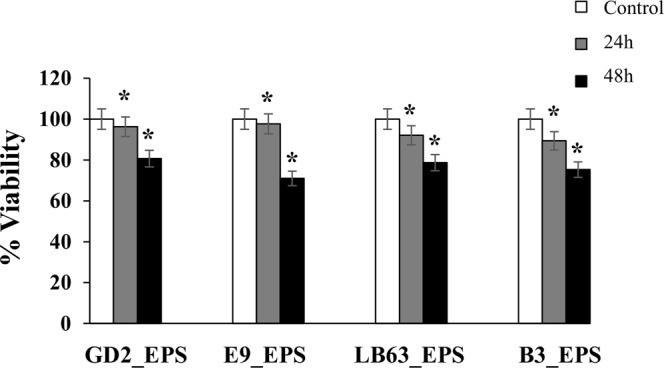
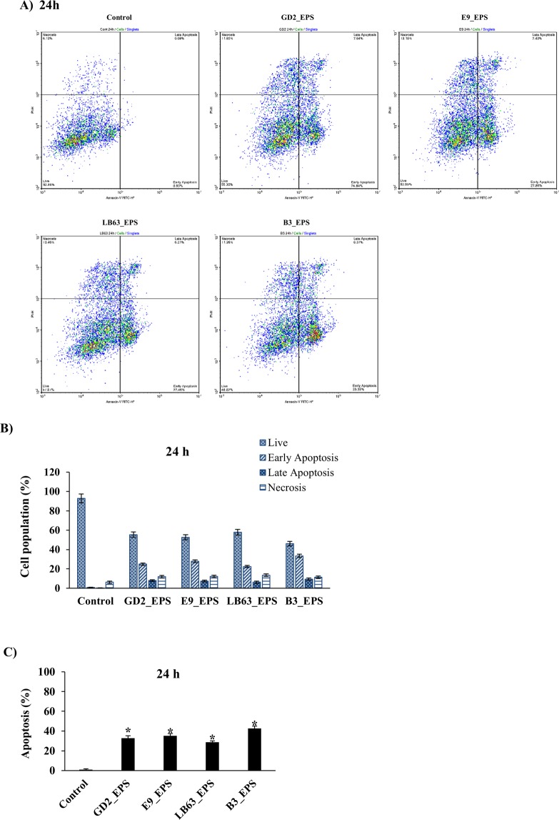
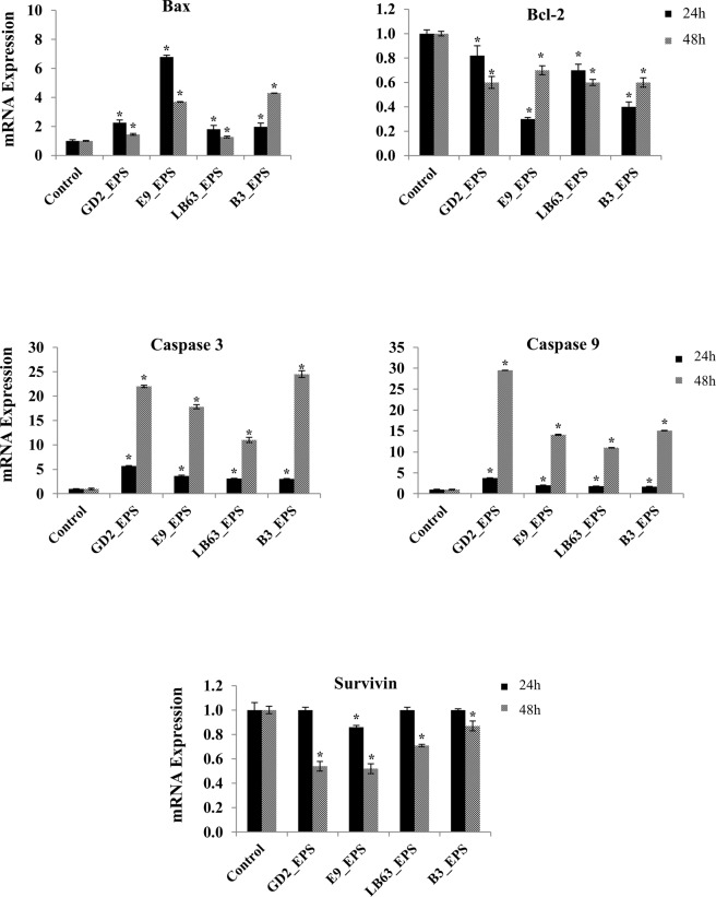
Colon cancer is one of the most common cancer around the world. Exopolysaccharides (EPSs) produced by lactobacilli as potential prebiotics have been found to have an anti-tumor effect. In this study, lyophilized EPSs of four Lactobacillus spp. for their impact on apoptosis in colon cancer cells (HT-29) was evaluated using flow cytometry. The relationship between capability of a lactobacilli-EPS to induce apoptosis and their monosaccharide composition, molecular weight (MW), and linkage type was investigated by HPLC, SEC, and NMR, respectively. Changes in apoptotic-markers were examined by qPCR and Western Blotting. EPSs were capable of inhibiting proliferation in a time-dependent manner and induced apoptosis via increasing the expression of Bax, Caspase 3 and 9 while decreasing Bcl-2 and Survivin. All EPSs contained mannose, glucose, and N-acetylglucosamine with different relative proportions. Some contained arabinose or fructose. MW ranged from 102-104Da with two or three fractions. EPS of L. delbrueckii ssp. bulgaricus B3 having the highest amount of mannose and the lowest amount of glucose, showed the highest apoptosis induction. In conclusion, lactobacilli-EPSs inhibit cell proliferation in HT-29 via apoptosis. Results suggest that a relationship exists between the ability of EPS to induce apoptosis and its mannose and glucose composition.


Conflict of interest statement

The authors declare no competing interests.

Figures

Figure 1
Figure 1
Repeating unit structure of the exopolysaccharides produced by Lactobacillus spp. NMR chemical shifts were determined by performing 1H-NMR, COSY and NOESY NMR analysis for the binding stereochemistry of monosaccharide units of EPSs of Lactobacillus spp. (L. plantarum GD2, L. rhamnosus E9, L. brevis LB63, and L. delbrueckii ssp. bulgaricus B3). All of them produced EPS with the same type of sugar linkage which are β-H1-H3 (β-D-Mannose-α-D-Mannose)/α- H1-H2 (α-D-Mannose-α-D-Glucose), designated as β-1,3 (β-D-Mannose-α-D-Mannose) and α-1,2 (α-D-Mannose-α-D-Glucose), respectively.
Figure 2
Figure 2
Anti-proliferative effect of the EPSs produced by Lactobacillus spp. against HT-29 colon cancer cells at two time points. HT-29 cells seeded at a density of 1 × 104 cells/well were treated with EPSs of Lactobacillus spp. (L. plantarum GD2, L. rhamnosus E9, L. brevis LB63, and L. delbrueckii ssp. bulgaricus B3) at a final concentration of 400 µg/ml for 24 h or 48 h and the anti-cytotoxicity effect of the EPSs was evaluated by WST-1 assay. *p < 0.05, significant difference from the control (n:3 for each bar).
Figure 3
Figure 3
Flow cytometric analysis of the impact of Lactobacillus spp. EPSs on apoptosis in HT-29 cells at 24 h time point. HT-29 cells seeded at a density of 1 × 104 cells/well were treated with EPSs of Lactobacillus spp. (L. plantarum GD2, L. rhamnosus E9, L. brevis LB63, and L. delbrueckii ssp. bulgaricus B3) at a final concentration of 400 µg/ml for 24 h and then stained with Annexin V-FITC and PI. Fluorescence intensities were detected by flow cytometry to determine the effect of the EPSs on earlier apoptosis, late apoptosis and necrosis. (A) Distribution of viable, early apoptotic, late apoptotic and necrotic cells analyzed by flow cytometry. (B) Percentage of cells in viable, early apoptotic, late apoptotic and necrotic stages. (C) Percentage of apoptosis in HT-29 cells exposed to the lactobacilli EPSs for 24 h. *p < 0.05, significant difference from the control.
Figure 4
Figure 4
Flow cytometric analysis of the impact of Lactobacillus spp. EPSs on apoptosis in HT-29 cells at 48 h time point. HT-29 cells seeded at a density of 1 × 104 cells/well were treated with EPSs of Lactobacillus spp. (L. plantarum GD2, L. rhamnosus E9, L. brevis LB63, and L. delbrueckii ssp. bulgaricus B3) at a final concentration of 400 µg/ml for 48 h and then stained with Annexin V-FITC and PI. Fluorescence intensities were detected by flow cytometry to determine the effect of the EPSs on earlier apoptosis, late apoptosis and necrosis. (A) Distribution of viable, early apoptotic, late apoptotic and necrotic cells analyzed by flow cytometry. (B) Percentage of cells in viable, early apoptotic, late apoptotic and necrotic stages. (C) Percentage of apoptosis in HT-29 cells exposed to the lactobacilli EPSs for 48 h. *p < 0.05, significant difference from the control.
Figure 5
Figure 5
Fold change in mRNA expression of target genes of the HT-29 cells in the control group and the group treated with EPSs from Lactobacillus spp. (L. plantarum GD2, L. rhamnosus E9, L. brevis LB63, and L. delbrueckii ssp. bulgaricus B3) at a final concentration of 400 µg/ml for 24 h (black bar) or 48 h (gray bar). *p < 0.05, significant difference from the control (n: 3).
Figure 6
Figure 6
Protein expression of target genes of the HT-29 cells in the control group and the group treated with EPSs from Lactobacillus spp. (L. plantarum GD2, L. rhamnosus E9, L. brevis LB63, and L. delbrueckii ssp. bulgaricus B3) at a final concentration of 400 µg/ml for 24 h. *p < 0.05, significant difference from the control (n:3).

References

    1. The International Agency for Research on Cancer (IARC). Latest Global Cancer Data, 2018. World Health Organization (2018).
    1. Bray F, et al. Global Cancer Statistics 2018: GLOBOCAN Estimates of Incidence and Mortality Worldwide for 36 Cancers in 185 Countries. CA Cancer J. Clin. 2018;68:394–424. doi: 10.3322/caac.21492. - DOI - PubMed
    1. Hill C, et al. The International Scientific Association for Probiotics and Prebiotics consensus statement on the scope and appropriate use of the term probiotic. Nat. Rev. Gastroenterol. Hepatol. 2014;11:506–514. doi: 10.1038/nrgastro.2014.66. - DOI - PubMed
    1. Gueimonde M, Ouwehand A, Huhtinen H, Salminen E, Salminen S. Qualitative and quantitative analyses of the bifidobacterial microbiota in the colonic mucosa of patients with colorectal cancer, diverticulitis and inflammatory bowel disease. World J Gastroenterol. 2007;13:3985–3989. doi: 10.3748/wjg.v13.i29.3985. - DOI - PMC - PubMed
    1. Kamada N, Núñez G. Regulation of the immune system by the resident intestinal bacteria. Gastroenterology. 2014;146:1477–88. doi: 10.1053/j.gastro.2014.01.060. - DOI - PMC - PubMed

Publication types

MeSH terms