. 2020 Jan;27(1):297-309.
doi: 10.1038/s41418-019-0358-x. Epub 2019 Jun 4.

Siva plays a critical role in mouse embryonic development

Affiliations

Siva plays a critical role in mouse embryonic development

Suzanne B R Jacobs et al. Cell Death Differ. 2020 Jan.

Abstract

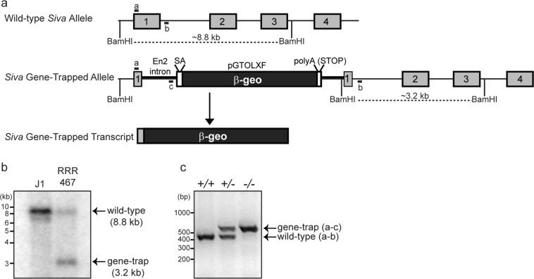
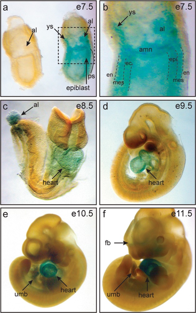
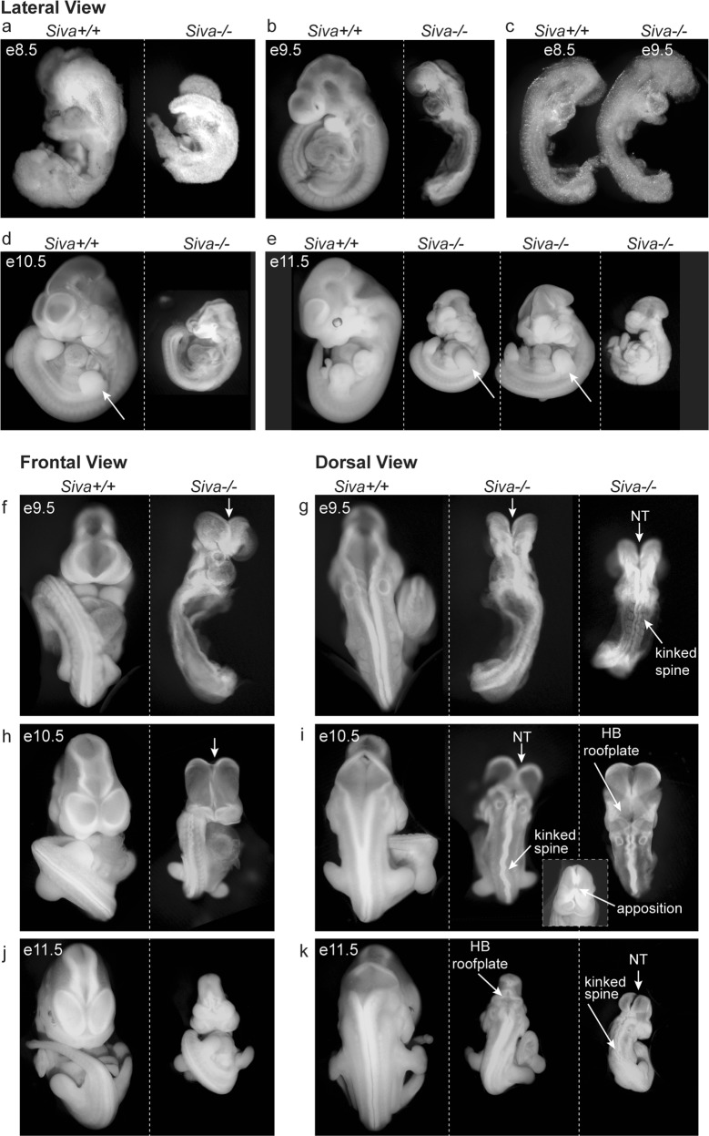
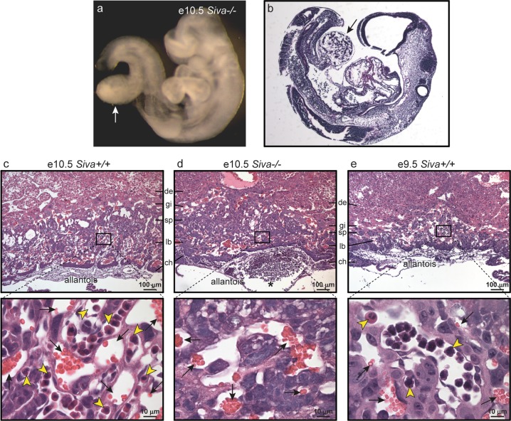
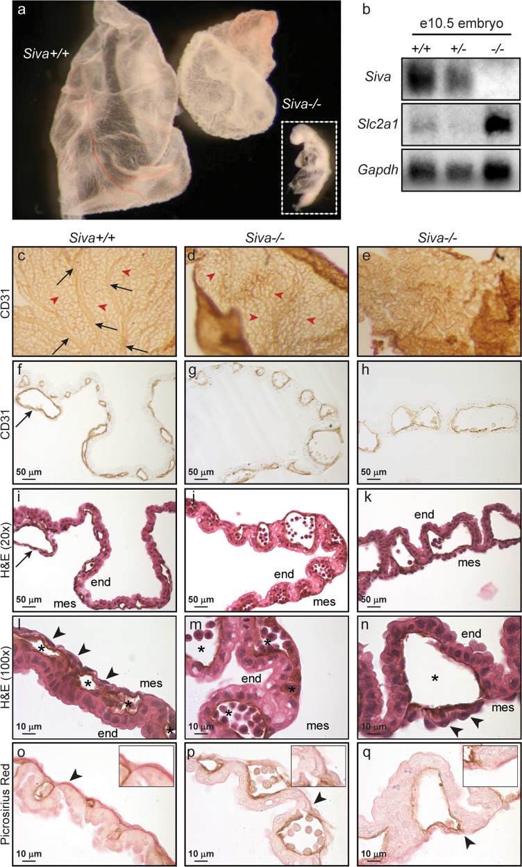
The Siva protein, named after the Hindu God of Destruction, plays important roles in apoptosis in various contexts, including downstream of death receptor activation or p53 tumor suppressor engagement. The function of Siva in organismal development and homeostasis, however, has remained uncharacterized. Here, we generate Siva knockout mice to characterize the physiological function of Siva in vivo. Interestingly, we find that Siva deficiency causes early embryonic lethality accompanied by multiple phenotypes, including developmental delay, abnormal neural tube closure, and defective placenta and yolk sac formation. Examination of Siva expression during embryogenesis shows that Siva is expressed in both embryonic and extra-embryonic tissues, including within the mesoderm, which may explain the vascular defects observed in the placenta and yolk sac. The embryonic phenotypes caused by Siva loss are not rescued by p53 deficiency, nor do they resemble those of p53 null embryos, suggesting that the embryonic function of Siva is not related to the p53 pathway. Moreover, loss of the Ripk3 necroptosis protein does not rescue the observed lethality or developmental defects, suggesting that Siva may play a non-apoptotic role in development. Collectively, these studies reveal a key role for Siva in proper embryonic development.


Conflict of interest statement

The authors declare that they have no conflict of interest.

Figures

Fig. 1
Fig. 1
Generation of Siva-knockout mice. a Schematic representation of Siva gene-trapped allele and transcript. The gene trap vector consists of Engrailed 2 (En2) intronic sequences followed by a splice acceptor (SA) and coding sequences for the β-galactosidase-neomycin (β-geo) gene. An SV40 polyA sequence acts as a transcriptional stop element. The gene-trapped transcript consists of the Siva 5’UTR and sequences encoding the first 3 amino acids from Siva exon1 fused to β-geo. BamHI sites and PCR primers used for genotyping are indicated. b Southern blot analysis of J1 (WT) and RRR467 (Siva gene trap) ES cell DNA demonstrates disruption of the Siva locus in the gene-trapped ES cells. Following DNA digestion with BamHI, Siva exon 2 sequences were used as a probe. (c) Genotyping PCR used to identify Siva+/+, Siva+/−, and Siva−/− mice and embryos. The forward primer recognizes a portion of 5′-UTR from Siva exon 1 (primer “a”) and is used in combination with reverse primers to a region of intron 1 (primer “b”) and the gene trap vector (primer “c”) The expected band sizes for the wild-type and gene-trapped alleles are 415 and 550 bp, respectively
Fig. 2
Fig. 2
Siva is expressed in embryonic and extra-embryonic tissues. a, b Whole-mount β-galactosidase staining of wild-type and Siva-heterozygous embryos at embryonic day e7.5 reveals Siva expression in the primitive streak, allantois, and yolk sac. b Higher magnification of box in (a) showing β-galactosidase expression in the primitive streak. (cf) Whole-mount β-galactosidase staining of embryos aged between e8.5 and e11.5 reveals continuing expression of Siva in the allantois and umbilical cord as well as in the heart and forebrain. al allantois, amn amnion, en endoderm, epi epiblast, fb forebrain, mes mesoderm, ec ectoderm, ps primitive streak, ys yolk sac, umb umbilical cord
Fig. 3
Fig. 3
Siva deficiency results in developmental delay and neurulation defects. ae Lateral view of morphology of wild-type and Siva−/− embryos from e8.5 through e11.5 reveals a developmental delay in the absence of Siva. Note that Siva−/− embryos are smaller and less developed than their littermates at all ages examined. Examination of limb development (arrows) demonstrates that Siva−/− embryos are developmentally delayed by ~1 day. In addition, quantitation of somite number in e9.5 Siva−/− embryos suggests a 1 day developmental delay. In (c), a side-by-side comparison of an e8.5 wild-type embryo with an e9.5 Siva−/− embryo demonstrates the similarity in size and developmental features, including the extent of neural tube closure, somite number, turning stage, and heart tube development. 54/57 e9.5–e11.5 Siva−/− embryos exhibited developmental delay. fk Morphological examination of neurulation defects (arrows) in Siva−/− embryos. Ventral and dorsal views of wild-type and Siva−/− embryos at e9.5 (f, g), e10.5 (h, i), and e11.5 (j, k). At all ages examined, Siva−/− embryos exhibit a failure to complete cranial neural tube closure (NT), kinked spines, and abnormally compacted hindbrains (HB)
Fig. 4
Fig. 4
Siva is required for chorioallantoic fusion and placental labyrinth formation. a Whole-mount image of e10.5 Siva−/− embryo in which chorioallantoic fusion failed to occur. The allantois (arrow) can be seen as a sac protruding from the embryonic gut. b H&E staining of embryo in (a), with arrow indicating allantois. ce Upper panels: Histological analysis of placentas from e9.5 and e10.5 embryos reveals defective labyrinth formation in the absence of Siva. H&E staining of placentas, in which chorioallantoic attachment occurred, shows that the Siva−/− allantois attaches but fails to penetrate the chorion. Lower panels: Higher magnification of boxed area of labyrinth layer. Embryonic and maternal blood vessels can be identified by the presence of nucleated erythroblasts (yellow arrowheads) and enucleated red blood cells (black arrows), respectively. Note the absence of embryonic blood vessels in the labyrinth from Siva−/− placentas (d). de decidua, gi trophoblast giant cells, sp spongiotrophoblast, lb labyrinth, ch chorion
Fig. 5
Fig. 5
Siva-knockout yolk sacs fail to undergo vascular remodeling. a Whole-mount examination of wild-type and Siva−/− e10.5 yolk sacs. Inset shows embryo associated with Siva−/− yolk sac. b Northern blot examining expression of the hypoxia marker Slc2a1 in Siva-null embryos. ce Whole-mount CD31 immunohistochemistry on wild-type and Siva−/− yolk sacs. The Siva−/− yolk sac shown in (e) is the same as shown in (a). fh Cross-sections of CD31-immunostained yolk sacs shown in (ce). ik H&E staining of CD31-immunostained yolk sacs. (ck) Arrows (black) indicate large vitelline vessels in wild-type yolk sac. Arrowheads (red) indicate small capillaries. ln Higher magnification of H&E-stained yolk sacs. Arrowheads indicate mesoderm cells. Note the loosely attached mesoderm cells in the Siva−/− yolk sac shown in (n) and the apparent absence of mesoderm cells in the Siva−/− yolk sac shown in (m). Asterisks denote endothelial cell-lined blood vessels. oq Picrosirius Red staining of yolk sac collagen (arrowheads) reveals a disruption in extracellular matrix production in the absence of Siva. end, endoderm; mes, mesoderm

References

    1. Prasad KV, Ao Z, Yoon Y, Wu MX, Rizk M, Jacquot S, et al. CD27, a member of the tumor necrosis factor receptor family, induces apoptosis and binds to Siva, a proapoptotic protein. Proc Natl Acad Sci USA. 1997;94:6346–51. - PMC - PubMed
    1. Spinicelli S, Nocentini G, Ronchetti S, Krausz LT, Bianchini R, Riccardi C. GITR interacts with the pro-apoptotic protein Siva and induces apoptosis. Cell Death Differ. 2002;9:1382–4. - PubMed
    1. Gudi R, Barkinge J, Hawkins S, Chu F, Manicassamy S, Sun Z, et al. Siva-1 negatively regulates NF-kappaB activity: effect on T-cell receptor-mediated activation-induced cell death (AICD) Oncogene. 2006;25:3458–62. - PubMed
    1. Gudi R, Barkinge J, Hawkins S, Prabhakar B, Kanteti P. Siva-1 promotes K-48 polyubiquitination of TRAF2 and inhibits TCR-mediated activation of NF-kappaB. J Environ Pathol Toxicol Oncol. 2009;28:25–38. - PMC - PubMed
    1. Resch U, Schichl YM, Winsauer G, Gudi R, Prasad K, de Martin R. Siva1 is a XIAP-interacting protein that balances NFkappaB and JNK signalling to promote apoptosis. J Cell Sci. 2009;122(Pt 15):2651–61. - PubMed

Publication types

MeSH terms

Substances