

Review
doi: 10.1152/physiol.00002.2019.
Whole-Cell cAMP and PKA Activity are Epiphenomena, Nanodomain Signaling Matters
Affiliations
- PMID: 31165682
- PMCID: PMC6863374
- DOI: 10.1152/physiol.00002.2019
Item in Clipboard
Review
Whole-Cell cAMP and PKA Activity are Epiphenomena, Nanodomain Signaling Matters
Physiology (Bethesda).
.
Abstract
Novel targeted fluorescent biosensors provide key insights into very local nanodomains of cAMP and PKA activity, and how they respond differently to β-adrenergic activation in cardiac myocytes. This unique spatiotemporal detail in living cells is not available with biochemical measurements of total cellular cAMP and PKA, and provides unique physiological insights.
Figures
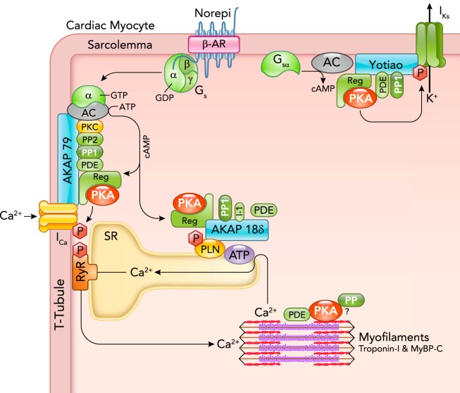
Cardiac myocyte β-AR, cAMP, and PKA signaling proteins are associated with AKAPs A β-AR activated by norepinephrine (Norepi) activates a GTPase protein (Gs) whose α-subunit activates adenylyl cyclase (AC) to produce cAMP from ATP. That cAMP binds to the regulatory subunit of PKA (Reg) to activate the catalytic subunit (shown as PKA) by relaxing the interaction with Reg. That active PKA can selectively phosphorylate nearby target proteins including sarcolemmal L-type Ca channels, which increases ICa, SR phospholamban (PLN), which increases SR Ca-ATPase activity, and KCNQ1 to enhance delayed rectifier K+ current IKs. PKA is anchored at these three targets, respectively, by AKAP79 (in human or mouse AKAP150; made by AKAP5 gene), AKAP18δ (made by AKAP7 gene), and Yotiao (made by AKAP9 gene). These AKAPs also bind additional cAMP-PKA modulators (PP1, PP2, PDEs, PKC). Troponin T has been proposed to be an AKAP (56), and TPNI and MyBP-C phosphorylation are regulated by PKA, PPs, and PDEs, but those mechanisms are less well resolved. See Ref. for a comprehensive AKAP review.
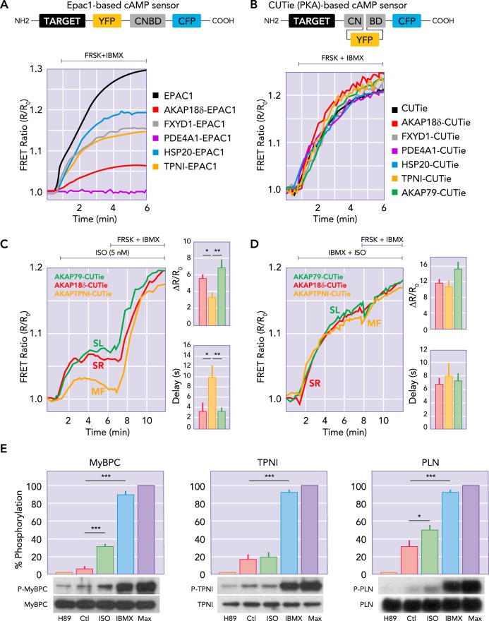
Targeted FRET-based reporters provide direct measurement of [cAMP] at discrete molecular sites in myocytes The more traditional cAMP reporter (A) has the cAMP binding domain (CNBD) between CFP and YFP, with the targeting domain (TARGET) attached to the NH2 terminal. The newly designed CUTie reporters (B) with different protein arrangement improves the consistency of the reporter affinity and dynamic range, regardless of targeting location (curves in B vs. A). In adult rat ventricular myocytes, targeted CUTies (C) indicated faster initial rise times and larger [cAMP] increases at the SL and SR vs. MF in response to 5 nM isoproterenol (ISO) exposure. When PDE were inhibited and AC was directly activated by forskolin, all three sensors gave the same signal (D), indicating that the nanodomain differences in [cAMP] induced by ISO were due in large part to the presence of PDE. E: PKA-dependent phosphorylation of MyBPC, TNI, and PLN at baseline (Ctl) in response to 0.3 nM ISO with minimum and maximum phosphorylation assessed by treatment with H89 (PKA inhibitor) or IBMX (or IBMX+forskolin; Max). Panels were redrawn based on parts of Figs. 1, 3, and 5 in Ref. , with permission from Nature Communications.

Key PKA targets downstream of β-AR activation in ventricular myocytes, their timing, and functional consequences PKA phosphorylates multiple target proteins in different domains [red hexagons (P)] at Ca2+, Na+, and K+ channels, SR Ca-ATPase and Na/K-ATPase (ATP), and myofilaments (TPNI, MyBP-C, and titin). Each site may be selectively tuned (both timing and kinetics) to optimize the orchestrated and integrated fight-or-flight response activated by the sympathetic nervous system. Table lists list tentative estimate of timing (numbers) and possibly strength, and related to Ca2+ handling (blue) or myofilament (brown).
References
-
- Barbagallo F, Xu B, Reddy GR, West T, Wang Q, Fu Q, Li M, Shi Q, Ginsburg KS, Ferrier W, Isidori AM, Naro F, Patel HH, Bossuyt J, Bers D, Xiang YK. Genetically encoded biosensors reveal PKA hyperphosphorylation on the myofilaments in rabbit heart failure. Circ Res 119: 931–943, 2016. doi:10.1161/CIRCRESAHA.116.308964. - DOI - PMC - PubMed
Publication types
MeSH terms
Substances
Grants and funding
LinkOut - more resources
Full Text Sources