. 2019 Jun 5;10(6):441.
doi: 10.1038/s41419-019-1631-0.

Mechanism of Snhg8/miR-384/Hoxa13/FAM3A axis regulating neuronal apoptosis in ischemic mice model

Affiliations

Mechanism of Snhg8/miR-384/Hoxa13/FAM3A axis regulating neuronal apoptosis in ischemic mice model

Jie Liu et al. Cell Death Dis. .

Erratum in

Abstract

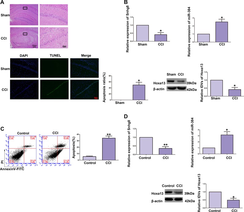
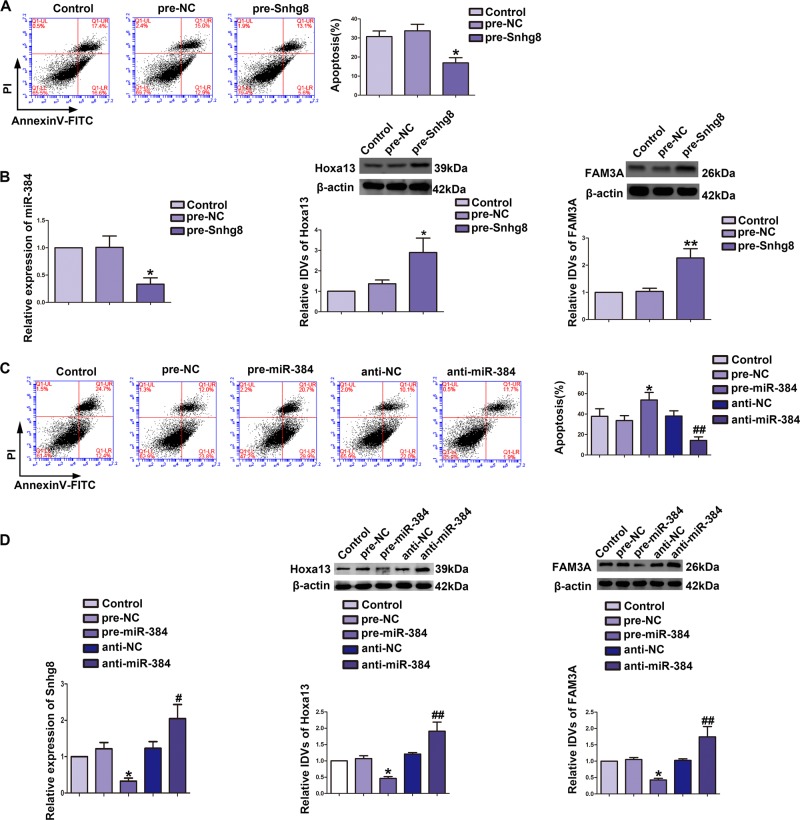
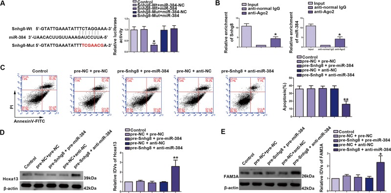
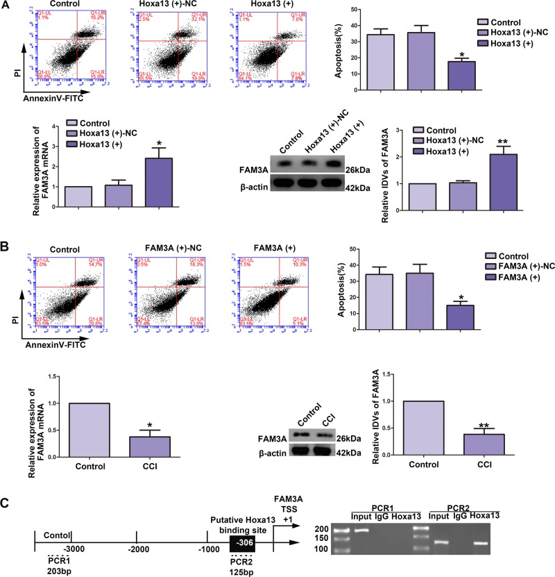
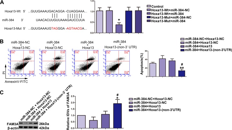
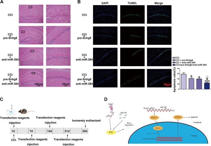
Long noncoding RNAs, a subgroup of noncoding RNAs, are implicated in ischemic brain injury. The expression levels of Snhg8, miR-384, Hoxa13, and FAM3A were measured in chronic cerebral ischemia-induced HT22 cells and hippocampal tissues. The role of the Snhg8/miR-384/Hoxa13/FAM3A axis was evaluated in chronic cerebral ischemia models in vivo and in vitro. In this study, we found that Snhg8 and Hoxa13 were downregulated, while miR-384 was upregulated in chronic cerebral ischemia-induced HT22 cells and hippocampal tissues. Overexpression of Snhg8 and Hoxa13, and silencing of miR-384, all inhibited chronic cerebral ischemia-induced apoptosis of HT22 cells. Moreover, Snhg8 bound to miR-384 in a sequence-dependent manner and there was a reciprocal repression between Snhg8 and miR-384. Besides, overexpression of miR-384 impaired Hoxa13 expression by targeting its 3'UTR and regulated chronic cerebral ischemia-induced neuronal apoptosis. Hoxa13 bound to the promoter of FAM3A and enhanced its promotor activity, which regulated chronic cerebral ischemia-induced neuronal apoptosis. Remarkably, the in vivo experiments demonstrated that Snhg8 overexpression combined with miR-384 knockdown led to an anti-apoptosis effect. These results reveal that the Snhg8/miR-384/Hoxa13/FAM3A axis plays a critical role in the regulation of chronic cerebral ischemia-induced neuronal apoptosis.


Conflict of interest statement

The authors declare that they have no conflict of interest.

Figures

Fig. 1
Fig. 1. Effect of chronic cerebral ischemia (CCI) to neuronal apoptosis and relative expression levels of Snhg8, miR-384, and Hoxa13.
a H&E-stained (upper part, ×100 or ×400, scale bar = 100 or 25 μm) and terminal deoxynucleotidyl transferase (TdT)-mediated dUTP-biotin nick end labeling (TUNEL) assay (lower part, ×200, scale bar = 50 μm) sections of the hippocampus CA1 region of Sham and CCI group (n = 4, each group). Data are represented as mean ± SD, *P < 0.05 vs. Sham group. b Quantitative real-time PCR and western blot (WB) were used to detect the relative expression levels of Snhg8 (upper left part, n = 5, each group), miR-384 (upper right part, n = 5, each group), and Hoxa13 (lower right part, n = 5, each group). Data are represented as mean ± SD, *P < 0.05 vs. Sham group. c Flow cytometry analysis of CCI-induced HT22 cells (n = 5, each group). Data are represented as mean ± SD, **P < 0.01 vs. Control group. d The expression levels of Snhg8 RNA (upper left part, n = 5, each group), miR-384 RNA (upper right part, n = 6, each group), and Hoxa13 protein (lower part, n = 5, each group) in CCI-induced HT22 cells. Data are represented as mean ± SD, *P < 0.05 vs. Control group, **P < 0.01 vs. Control group
Fig. 2
Fig. 2. Snhg8 protected chronic cerebral ischemia (CCI)-induced neuronal apoptosis, while miR-384 exacerbated CCI-induced neuronal apoptosis.
a Flow cytometry analysis of CCI-induced HT22 cells with overexpression of Snhg8 (n = 5, each group). Data are represented as mean ± SD, *P < 0.05 vs. Pre-NC group. b Expression analysis of miR-384 RNA (left part, n = 5, each group), Hoxa13 protein (middle part, n = 5, each group), and FAM3A protein (right part, n = 5, each group) in CCI-induced HT22 cells. Data are represented as mean ± SD, *P < 0.05 vs. Pre-NC group, **P < 0.01 vs. Pre-NC group. c Flow cytometry analysis of CCI-induced HT22 cells with transfection of miR-384 (n = 4, each group). Data are represented as mean ± SD, *P < 0.05 vs. Pre-NC group, ##P < 0.01 vs. anti-NC group. d Expression analysis of Snhg8 RNA (left part, n = 5, each group), Hoxa13 protein (middle part, n = 4, each group) and FAM3A protein (right part, n = 6, each group) in CCI-induced HT22 cells with transfection of miR-384. Data are represented as mean ± SD, *P < 0.05 vs. Pre-NC group, #P < 0.05 vs. anti-NC group, ##P < 0.01 vs. anti-NC group
Fig. 3
Fig. 3. miR-384-targeted Snhg8 and reversed the Snhg8-mediated attenuation of chronic cerebral ischemia (CCI)-induced neuronal apoptosis.
a The predicted miR-384-binding site in Snhg8 (Snhg8-Wt) and the designed mutant sequence (Snhg8-Mut) were indicated. Luciferase reporter assay of HT22 cells cotransfected with Snhg8-Wt or Snhg8-Mut and miR-384 or the miR-384-NC (data are presented as the mean ± SD (n = 4, each group). Data are displayed as mean ± SD, *P < 0.05 vs. Snhg8-Wt+miR-384-NC group. b miR-384 was identified in Snhg8-RISC complex. Relative expression levels of Snhg8 (n = 4, each group) and miR-384 were determined by qRT PCR (n = 4, each group). Data are displayed as mean ± SD, *P < 0.05 vs. anti-normal IgG. c Flow cytometry analysis of CCI-induced HT22 cells cotransfected with Snhg8 and miR-384 (n = 4, each group). Data are displayed as mean ± SD, **P < 0.01 vs. pre-NC+anti-NC group. d Hoxa13 protein levels in CCI-induced HT22 cells cotransfected with Snhg8 and miR-384 (n = 4, each group). Data are displayed as mean ± SD, **P < 0.01 vs. pre-NC+anti-NC group. e FAM3A protein levels in CCI-induced HT22 cells cotransfected with Snhg8 and miR-384 (n = 4, each group). Data are displayed as mean ± SD, **P < 0.01 vs. pre-NC+anti-NC group
Fig. 4
Fig. 4. Overexpression of Hoxa13 and overexpression of FAM3A inhibited chronic cerebral ischemia (CCI) induced neuronal apoptosis.
a Flow cytometry analysis of CCI-induced HT22 cells with overexpression of Hoxa13 (upper part, n = 4, each group). Expression analysis of FAM3A RNA (lower left part, n = 5, each group) and FAM3A protein (lower right part, n = 5, each group) in CCI-induced HT22 cells transfected with Hoxa13. Data are represented as mean ± SD, *P < 0.05 vs. Hoxa13 (+)-NC group, **P < 0.01 vs. Hoxa13 (+)-NC group. b Flow cytometry analysis of chronic cerebral ischemia CCI-induced HT22 cells with overexpression of FAM3A (upper part, n = 3, each group). Expression analysis of FAM3A RNA (lower left part, n = 4, each group) and FAM3A protein (lower right part, n = 6, each group) in CCI-induced HT22 cells. Data are represented as mean ± SD, *P < 0.05 vs. FAM3A (+)-NC group, *P < 0.05 vs. Control group, **P < 0.01 vs. Control group. c Putative FAM3A-binding sites are indicated. Immunoprecipitated DNA was amplified by PCR. Normal rabbit IgG was used as a negative control
Fig. 5
Fig. 5. Overexpression of miR-384 impaired Hoxa13-induced attenuation of chronic cerebral ischemia (CCI)-induced neuronal apoptosis by targeting 3´-UTR of Hoxa13.
a The predicted miR-384-binding site in Hoxa13 and the designed mutant sequences were indicated. Dual-luciferase report assay of HT22 cells cotransfected with Hoxa13-Wt and miR-384-NC or Hoxa13-Wt and miR-384, and Hoxa13-Mut and miR-384-NC or Hoxa13-Mut and miR-384 (n = 4, each group). Data are presented as mean ± SD, *P < 0.05 vs. Hoxa13-Wt+miR-384-NC group. b Flow cytometry analysis of CCI-induced HT22 cells cotransfected with miR-384 and Hoxa13/Hoxa13- (non-3´UTR) (n = 4, each group). Data are presented as mean ± SD, **P < 0.01 vs. miR-384+Hoxa13-NC group, #P < 0.05 vs. miR-384+Hoxa13 group. c FAM3A protein levels in CCI-induced HT22 cells cotransfected with miR-384 and Hoxa13/Hoxa13-(non-3´UTR) (n = 4, each group). Data are presented as mean ± SD, *P < 0.05 vs. miR-384+Hoxa13-NC group, #P < 0.05 vs. miR-384+Hoxa13 group
Fig. 6
Fig. 6. Overexpression of Snhg8 combined with knockdown of miR-384 protected against chronic cerebral ischemia (CCI) induced neuronal apoptosis in vivo.
a H&E-stained (×100, ×400, scale bar = 100 and 25 μm) and b terminal deoxynucleotidyl transferase (TdT)-mediated dUTP-biotin nick end labeling (TUNEL) assay (×200, scale bar = 50 μm) 0f CA1 area of hippocampus tansfected with Snhg8 and miR-384 in vivo (n = 5, each group). Data are presented as mean ± SD. *P < 0.05 vs. CCI group, ***P < 0.001 vs. CCI group, #P < 0.05 vs. CCI+pre-Snhg8, △P < 0.05 vs. CCI+miR-384 group. c Experimental schedule to explore the effects of Snhg8 and miR-384 on CCI-induced neuronal damage in vivo. d schematic illustration of the mechanism of Snhg8/miR-384/ Hoxa13/FAM3A axis regulating neuronal apoptosis

Similar articles

Cited by

References

    1. Farkas E, Luiten PG. Cerebral microvascular pathology in aging and Alzheimer’s disease. Prog. Neurobiol. 2001;64:575–611. doi: 10.1016/S0301-0082(00)00068-X. - DOI - PubMed
    1. Farkas E, Luiten PG, Bari F. Permanent, bilateral common carotid artery occlusion in the rat: a model for chronic cerebral hypoperfusion-related neurodegenerative diseases. Brain Res. Rev. 2007;54:162–180. doi: 10.1016/j.brainresrev.2007.01.003. - DOI - PubMed
    1. Bennett SA, et al. Chronic cerebral hypoperfusion elicits neuronal apoptosis and behavioral impairment. Neuroreport. 1998;9:161–166. doi: 10.1097/00001756-199801050-00033. - DOI - PubMed
    1. Bink DI, Ritz K, Aronica E, van der Weerd L, Daemen MJ. Mouse models to study the effect of cardiovascular risk factors on brain structure and cognition. J. Cereb. Blood Flow Metab. 2013;33:1666–1684. doi: 10.1038/jcbfm.2013.140. - DOI - PMC - PubMed
    1. Shibata M, Ohtani R, Ihara M, Tomimoto H. White matter lesions and glial activation in a novel mouse model of chronic cerebral hypoperfusion. Stroke. 2004;35:2598–2603. doi: 10.1161/01.STR.0000143725.19053.60. - DOI - PubMed

Publication types

MeSH terms