. 2019 Jun 5;51(6):1-16.
doi: 10.1038/s12276-019-0264-9.

Metformin treatment reduces motor and neuropsychiatric phenotypes in the zQ175 mouse model of Huntington disease

Affiliations

Metformin treatment reduces motor and neuropsychiatric phenotypes in the zQ175 mouse model of Huntington disease

Ana Sanchis et al. Exp Mol Med. .

Abstract

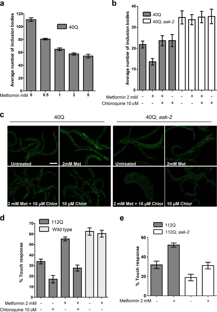
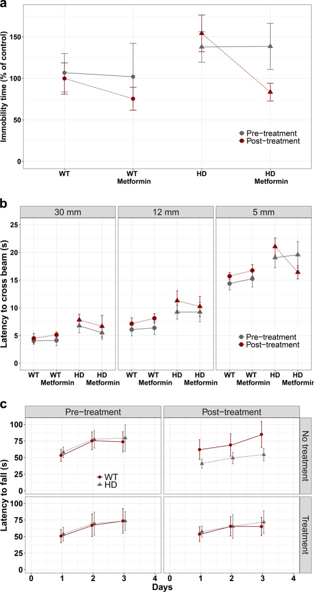
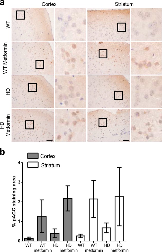
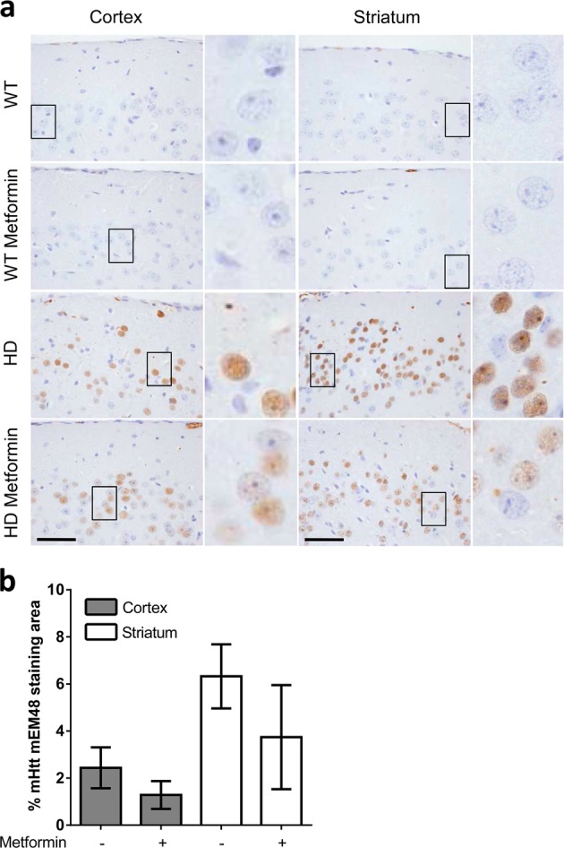
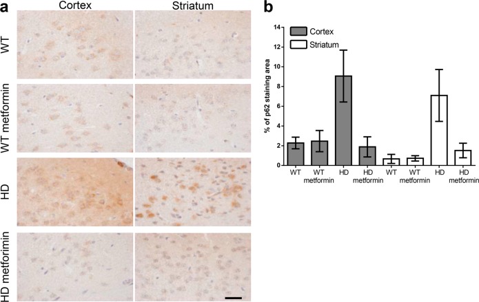
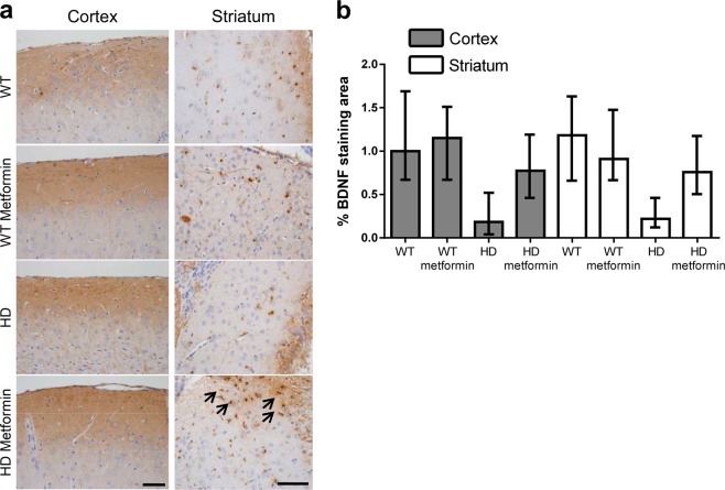
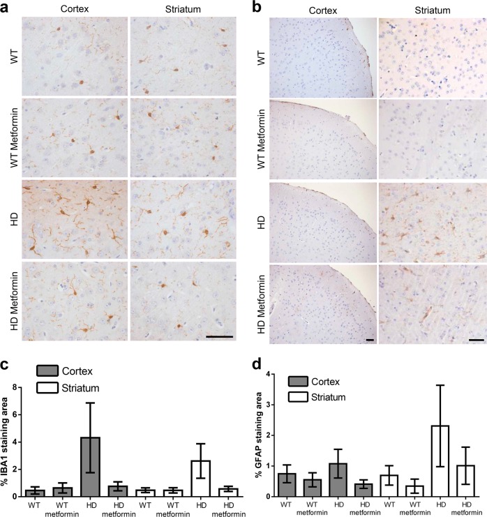
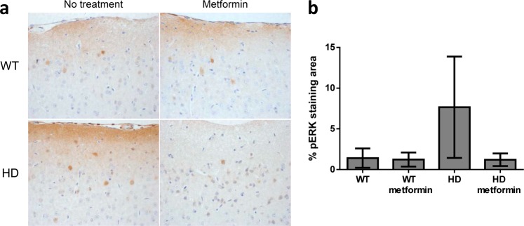
Huntington disease is a neurodegenerative condition for which there is no cure to date. Activation of AMP-activated protein kinase has previously been shown to be beneficial in in vitro and in vivo models of Huntington's disease. Moreover, a recent cross-sectional study demonstrated that treatment with metformin, a well-known activator of this enzyme, is associated with better cognitive scores in patients with this disease. We performed a preclinical study using metformin to treat phenotypes of the zQ175 mouse model of Huntington disease. We evaluated behavior (motor and neuropsychiatric function) and molecular phenotypes (aggregation of mutant huntingtin, levels of brain-derived neurotrophic factor, neuronal inflammation, etc.). We also used two models of polyglutamine toxicity in Caenorhabditis elegans to further explore potential mechanisms of metformin action. Our results provide strong evidence that metformin alleviates motor and neuropsychiatric phenotypes in zQ175 mice. Moreover, metformin intake reduces the number of nuclear aggregates of mutant huntingtin in the striatum. The expression of brain-derived neurotrophic factor, which is reduced in mutant animals, is partially restored in metformin-treated mice, and glial activation in mutant mice is reduced in metformin-treated animals. In addition, using worm models of polyglutamine toxicity, we demonstrate that metformin reduces polyglutamine aggregates and restores neuronal function through mechanisms involving AMP-activated protein kinase and lysosomal function. Our data indicate that metformin alleviates the progression of the disease and further supports AMP-activated protein kinase as a druggable target against Huntington's disease.


Conflict of interest statement

The authors declare that they have no conflict of interest.

Figures

Fig. 1
Fig. 1. PolyQ toxicity in C. elegans is reduced upon AMP-activated protein kinase (AMPK) activation and requires lysosome function.
a Metformin was able to reduce aggregation in a dose-dependent manner in 1-day-old adult worms expressing 40Q::YFP in muscle cells. Treatment with metformin significantly rescued aggregates in a dose-dependent manner (p < 0.001). b Metformin required aak-2/AMPKα and lysosome function to reduce the number of inclusion bodies in L4 larvae. Larvae were used to test for AMPK function because the difference between mutant and wild-type worms is reduced as the animals age (data not shown). Treatment with metformin rescued aggregates in 40Q animals (p < 0.001). Chloroquine maintained high levels of inclusion bodies even when the 40Q worms are treated with metformin (p < 0.001). Metformin required aak-2/AMPK to partially rescue polyQ aggregation (p < 0.001). c Photographs of wild-type and aak-2/AMPKα mutant L4 larvae expressing 40Q::YFP in muscle cells treated with or without 2 mM metformin and/or 10 μM chloroquine. Larvae were used to test for AMPK function because the difference between mutant and wild-type worms is reduced as the animals age (data not shown). d Treatment with metformin significantly rescued neuronal function (assessed by the touch response; see Materials and methods) in 112Q-TdTomato worms (p < 0.001). The metformin-induced rescue of neuronal function required lysosomal function: chloroquine treatment reduced neuronal recovery even when the worms were treated with metformin (p = 0.261). e Worms require AMPKα activity to rescue the neuronal function induced by metformin. Metformin was applied to worms expressing 112Q-TdTomato in mechanosensory neurons in an aak-2 background. The touch response was assessed as above. Metformin required aak-2/AMPK to rescue neuronal function (p = 0.634). In all cases, values are the mean, and bars indicate the confidence interval (95% CI)
Fig. 2
Fig. 2. Metformin treatment ameliorates the neuropsychiatric and motor behavior phenotype in heterozygous zQ175 mice.
Different behavioral tests were analyzed in 3-month-old mice (pretreatment) and after 3 months of treatment (6-month-old; post treatment). a Huntington disease (HD) and control mice were subjected to the tail suspension test, and the immobility time was measured (see Materials and methods). HD mice showed a depressive state as early as 3 months of age, which was worse in 6-month-old animals. However, when the mice were treated with metformin, their depression-related behavior became similar to that in wild-type (WT) animals (p < 0.037). b HD and control animals were subjected to the beam balance test. The time to cross each beam was recorded. HD mice exhibited difficulties crossing beams of different widths compared to WT animals (30 mm: p < 0.001; 12 mm: p < 0.001; and 5 mm: p < 0.001). In contrast, HD mice treated with metformin showed a reduced latency to cross the beam in comparison to non-treated mice (p = 0.014), and the values were similar to those for WT controls in the case of the 5 mm width beam (p = 0.822). c Rotarod experiments were performed with HD and control mice as described in the Materials and methods. Three-month-old HD animals had similar motor coordination to WT mice, although this motor behavior worsened with age (p < 0.001). However, when HD animals were treated with metformin, they showed higher latency to fall (p < 0.001), and they maintained a similar motor behavior to WT mice (p = 0.795). Values are the mean, and bars indicate the confidence interval (95% CI)
Fig. 3
Fig. 3. Levels of phospho-acetyl CoA carboxylase (pACC), a substrate of AMP-activated protein kinase (AMPK), in brain tissue of 6-month-old control and zQ175 mice treated or not with metformin.
a Immunohistochemical (IHC) staining illustrates the expression levels of pACC in the cortex and striatum of zQ175 (Huntington disease (HD)) mice and their corresponding controls (wild type (WT)). Representative images of each condition are shown (bars: 50 µm). Boxed areas are enlarged, with a higher magnification, on the right side of each image. b The bottom panel shows a semiquantitative analysis of IHC staining using the ImageJ software. Samples from four independent mice from each group were analyzed, which generated 7 cortex and 12 striatum pictures from untreated wild-type (WT) mice, 9 cortex and 12 striatum pictures from treated wild-type (WT metformin) mice, 7 cortex and 6 striatum pictures from untreated zQ175 (HD) mice, and 10 cortex and 6 striatum pictures from HD-treated (HD metformin) mice. The analyzed area in each picture was 90,488 μm2. Treated mice showed increased levels of pACC, independent of their genotype (p < 0.001). Values are the mean, and bars indicate the confidence interval (95% CI)
Fig. 4
Fig. 4. Mutant huntingtin aggregation in the brain tissue of six-month-old zQ175 mice is reduced by metformin treatment.
a Immunohistochemical (IHC) staining illustrates the expression levels of mutant huntingtin (mHTT) in the cortex and striatum of zQ175 mice and their corresponding controls (wild type (WT)). Representative images of each condition are shown (bars: 50 µm). Boxed areas are enlarged, with a higher magnification, on the right side of each image. b The bottom panel shows a semiquantitative analysis of IHC staining using the ImageJ software. Samples from four independent mice from each group were analyzed, which generated 10 cortex and 8 striatum pictures from untreated zQ175 (Huntington disease (HD)) mice and 10 cortex and 7 striatum pictures from HD-treated (HD metformin) mice. The analyzed area in each picture was 90,488 μm2. HD mice treated with metformin showed fewer mHtt aggregates (p = 0.00243), and the reduction in the aggregates was more pronounced in the striatum of these mice (p < 0.001). Values are the mean, and bars indicate the confidence interval (95% CI)
Fig. 5
Fig. 5. Levels of autophagy receptor p62 in brain tissue of 6-month-old control and zQ175 mice treated or not with metformin.
a Immunohistochemical (IHC) staining illustrates the expression levels of p62 in the cortex and striatum of zQ175 (Huntington disease (HD)) mice and their corresponding controls (wild type (WT)). Representative images of each condition are shown (bars: 50 µm). b Semiquantitative analysis of IHC staining using the ImageJ software. Samples from four independent mice from the WT and nontreated HD groups and 5 mice from the treated HD group were analyzed, which generated 13 cortex and 8 striatum pictures from untreated wild-type (WT), 12 cortex and 7 striatum pictures from treated wild-type (WT metformin), 14 cortex and 7 striatum pictures from untreated zQ175 (HD), and 14 cortex and 13 striatum pictures from HD-treated (HD metformin) mice. The analyzed area in each picture was 90,488 μm2. HD mice showed higher levels of p62 than WT mice (p < 0.001), and treatment with metformin reduced the levels of p62 (p < 0.001) to values similar to those in WT mice. Values are the mean, and bars indicate the confidence interval (95% CI)
Fig. 6
Fig. 6. Metformin prevents the loss of brain-derived neurotrophic factor (BDNF) levels in Huntington disease (HD) mice.
a Immunohistochemical (IHC) staining of the striatum of HD and control mice using anti-BDNF antibodies. Arrows denote the localization of BDNF in treated HD samples. Representative images of each condition are shown (bar: 50 µm). b Semiquantitative analysis of IHC staining using the ImageJ software. Samples from four independent mice from wild-type (WT) and nontreated HD groups and five mice from the treated HD group were analyzed, which generated 4 cortex and 4 striatum pictures from untreated wild-type (WT), 4 cortex and 4 striatum pictures from treated wild-type (WT metformin), 4 cortex and 4 striatum pictures from untreated zQ175 (HD), and 5 cortex and 5 striatum pictures from HD-treated (HD metformin) mice. The analyzed area in each picture was 90,488 μm2. The protein levels of BDNF in HD mice were lower than those in WT littermates (p < 0.001), and metformin treatment increased the protein levels (p < 0.001). HD mice treated with metformin showed similar levels of BDNF as WT littermates (p = 0.853). Values are the mean, and bars indicate the confidence interval (95% CI)
Fig. 7
Fig. 7. The inflammatory response in the brain tissue of 6-month-old zQ175 mice is reduced after metformin treatment.
Immunohistochemical (IHC) staining of the striatum of Huntington disease (HD) and control mice using anti-ionized calcium-binding adaptor molecule 1 (IBA1) (a) and anti-glial fibrillary acidic protein (GFAP) (b) antibodies. IHC staining of IBA1 and GFAP were significantly reduced by metformin treatment. Representative images of each condition are shown (bar: 50 µm). c, d The semiquantitative analysis of IHC staining of both biomarkers using the ImageJ software. The number of independent mice and samples from each is as follows: (1) for GFAP, we analyzed 4 nontreated wild type (WT), 3 treated WT, 4 nontreated HD, and 5 treated HD mice, which generated 10 cortex and 8 striatum pictures from untreated wild-type (WT), 6 cortex and 8 striatum pictures from treated wild-type (WT metformin), 4 cortex and 6 striatum pictures from untreated zQ175 (HD), and 7 cortex and 10 striatum pictures from HD treated (HD metformin) mice. The analyzed area in each picture was 361,920 μm2; (2) for IBA1, we analyzed 4 mice from each group (nontreated and treated WT mice and nontreated and treated HD mice), which generated 4 cortex and 10 striatum pictures from untreated wild-type (WT), 7 cortex and 5 striatum pictures from treated wild-type (WT metformin), 6 cortex and 9 striatum pictures from untreated zQ175 (HD) and 11 cortex and 13 striatum pictures from HD treated (HD metformin) mice. The analyzed area in each picture was 36,580 μm2. c Analysis of these data indicates that HD mice expressed higher levels of IBA1 than WT mice in both the cortex and striatum (p < 0.001 in both cases) and that metformin reduced the levels of IBA1 to those of WT mice (p = 0.932). d HD mice expressed higher levels of GFAP than WT mice (p < 0.001). Treatment with metformin significantly reduced GFAP levels in HD mice to levels similar to those of WT mice (p = 0.33). Values are the mean, and bars indicate the confidence interval (95% CI)
Fig. 8
Fig. 8. Metformin treatment reduces phospho-extracellular signal-regulated kinase1/2 (pERK1/2) expression in Huntington disease (HD) mice.
a Immunohistochemical (IHC) staining of the cortex of Huntington disease (HD) and control mice using the anti-pERK1/2 antibody. Representative images of each condition are shown (bar: 50 µm). b Semiquantitative analysis of IHC staining using the ImageJ software. Samples from four independent mice from each group, except for HD-treated mice, of which we used six mice, were analyzed. These mice generated the following material: 12 cortex pictures from untreated wild-type (WT), 12 cortex pictures from treated wild-type (WT metformin), 12 cortex pictures from untreated zQ175 (HD), and 18 cortex pictures from HD treated (HD metformin) mice. The analyzed area in each picture was 90,488 μm2. HD mice showed higher levels of pERK1/2 than WT mice (p = 0.011). Treatment with metformin reduced pERK1/2 levels (p = 0.013) to levels similar to those in WT mice (p = 0.686). Values are the mean, and bars indicate the confidence interval (95% CI)

Similar articles

Cited by

References

    1. Krobitsch S, Kazantsev AG. Huntington's disease: from molecular basis to therapeutic advances. Int. J. Biochem. Cell Biol. 2011;43:20–24. doi: 10.1016/j.biocel.2010.10.014. - DOI - PubMed
    1. Ferrante RJ, Kowall NW, Richardson EP., Jr Proliferative and degenerative changes in striatal spiny neurons in Huntington's disease: a combined study using the section-Golgi method and calbindin D28k immunocytochemistry. J. Neurosci. 1991;11:3877–3887. doi: 10.1523/JNEUROSCI.11-12-03877.1991. - DOI - PMC - PubMed
    1. Yamamoto A, Cremona ML, Rothman JE. Autophagy-mediated clearance of huntingtin aggregates triggered by the insulin-signaling pathway. J. Cell Biol. 2006;172:719–731. doi: 10.1083/jcb.200510065. - DOI - PMC - PubMed
    1. Ortega Z, Lucas JJ. Ubiquitin-proteasome system involvement in Huntington’s disease. Front. Mol. Neurosci. 2014;7:77. doi: 10.3389/fnmol.2014.00077. - DOI - PMC - PubMed
    1. Herrero-Martin G, et al. TAK1 activates AMPK-dependent cytoprotective autophagy in TRAIL-treated epithelial cells. EMBO J. 2009;28:677–685. doi: 10.1038/emboj.2009.8. - DOI - PMC - PubMed

Publication types

MeSH terms