. 2019 Jun 5;10(6):443.
doi: 10.1038/s41419-019-1686-y.

Deer antler stem cells are a novel type of cells that sustain full regeneration of a mammalian organ-deer antler

Affiliations

Deer antler stem cells are a novel type of cells that sustain full regeneration of a mammalian organ-deer antler

Datao Wang et al. Cell Death Dis. .

Abstract

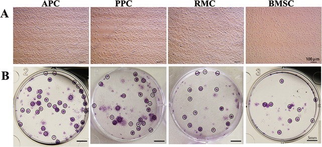
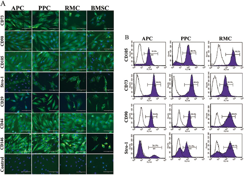
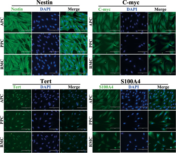
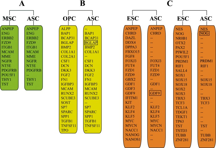
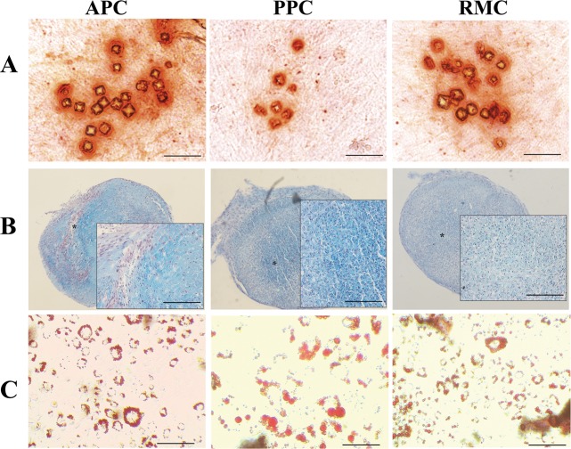
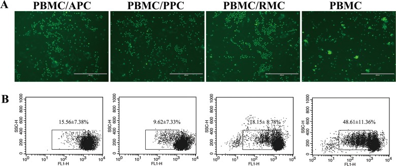
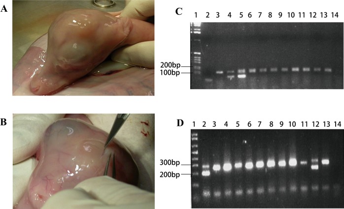
Deer antlers are extraordinary mammalian organs that can fully regenerate annually. Antler renewal is a stem cell-based epimorphic process and antler stem (AS) cells can initiate de novo generation of antlers in postnatal mammals. However, although being called stem cells, the AS cells have not been characterized at molecular level based on the stem cell criteria. Comprehensive characterization of the AS cells would undoubtedly help to decipher the mechanism underlying the full regeneration of deer antlers, the only case of stem cell-based epimorphic regeneration in mammals. In the present study, three types of AS cells (antlerogenic periosteal cells APCs, for initial pedicle and first antler formation; pedicle periosteal cells PPC, for annual antler regeneration; and reserve mesenchyme cells RMCs, for rapid antler growth), were isolated for comprehensive molecular characterization. A horn-growth-related gene, RXFP2, was found to be expressed only in AS cells lineages but not in the facial periosteal cells (FPCs, locates geographically in the vicinity of the APCs or PPCs), suggesting the RXFP2 might be a specific marker for the AS cell lineage in deer. Our results demonstrated that AS cells expressed classic MSC markers including surface markers CD73, CD90, CD105 and Stro-1. They also expressed some of the markers including Tert, Nestin, S100A4, nucleostemin and C-Myc, suggesting that they have some attributes of the ESCs. Microinjection of male APC into deer blastocysts resulted in one female foetus (110 days gestation) recovered with obvious pedicle primordia with both male and female genotype detected in the ovary. In conclusion, the AS cells should be defined as MSCs but with partial attributes of ESCs.


Conflict of interest statement

The authors declare that they have no conflict of interest.

Figures

Fig. 1
Fig. 1. Morphological observation and colony formation of AS cells.
a Cells from antler relative tissues were isolated and cultured as described in methodology section; their morphology was monitored under microscope (Passages ≤ 5); Bar = 100 μm. b Colonies formed after seeding 100 cells/well at day 14, and were stained with crystal violet dye; Bar = 5 mm; APC antlerogenic periosteal cell, PPC pedicle periosteal cell, RMC reserve mesenchymal cell, BMSC bone marrow stem cell
Fig. 2
Fig. 2. Expression of surface stem cell markers in AS cells.
a Immunofluorescence staining of AS cells. Classical surface markers of stem cells (CD73, CD90, CD105, Stro-1, CD29, CD44, and CD146) were detected using immunofluorescence staining (Green). Cell nuclei were counterstained with DAPI (Blue). Scale bar = 100 µm. b Flow cytometry analysis of AS cells. Expressions of indicated antigen are shown in purple histograms in contrast to isotype controls (black histograms). Values showed positive expression patterns of the indicated antigen
Fig. 3
Fig. 3. Immunofluorescence staining of intracellular markers in the AS cells.
Nestin, C-myc, Tert and S100A4 (known as intracellular markers) were detected using immunofluorescence staining. Cell nuclei were counterstained with DAPI (Blue). Note that filamentous Nestin distributed in the whole cytoplasm and Tert enriched in cell nuclei. Scale bar = 100 µm
Fig. 4
Fig. 4. Expression of stem cell markers in AS cells (ASC).
a Expression of mesenchymal stem cell (MSC) markers. b Expression of osteoprogenitor cells (OPC) markers. c: expression of embryonic stem cell (ESC) markers. The list of indicated stem cell markers were defined by Pazhanisamy (2013)
Fig. 5
Fig. 5. Multipotency of AS cells.
a Osteogenic differentiation - Alizarin Red S and Von Kossa staining after 3 weeks of osteogenic induction; differentiated cells stained strongly with Alizarin Red S. b Chondrogenic differentiation - chondrogenic pellets were cut into 5-mm sections for Alcian blue-PAS staining; the stained tissue displayed a typical cartilaginous tissue phenotype. c Adipogenic differentiation - Oil Red O staining was conducted after 2 weeks of adipogenic induction; AS cells exhibited Oil Red-O positive lipid droplets
Fig. 6
Fig. 6. AS cells significantly suppressed the proliferation of active lymphocytes.
a Lymphocytes were labelled with CFSE and co-cultured with AS cells at a ratio of 1:10 (ASC: lymphocyte) for 4 days and observed under fluorescence microscope; cell masses were proliferating lymphocytes; note that the co-cultured lymphocytes were almost devoid of cell masses. b Reduction of CFSE fluorescence intensity in PBMCs was analyzed by flow cytometry; data are expressed as the mean ± SD from three independent experiments
Fig. 7
Fig. 7. Production of chimera from APCs.
a Male fetus derived from blastocyst injected with morula embryo cells; neck girth of 9.0 cm. b Female fetus derived from blastocyst injected with male AP cells. Note the prominent pedicle formation on the head; neck girth of 9.5 cm. c PCR amplification of SRY region of the Y chromosome (male 104 bp) and internal DNA positive control (194 bp) of various tissues from the female fetus with pedicles. (1) Ladder; (2) Water control; (3) Female control; (4) Male control; (5) Ovary (male DNA in the ovary); (6) Kidney; (7) Skin; (8) Pedicles; (9) Skin pedicles; (10) Heart; (11) Gut; (12) Brainstem; (13) Muscle; (14) Blood. d PCR amplification of amelogenin gene (male sequence deletion 200 bp) and female 300 bp of various tissues from the female fetus with pedicles. (1) Ladder; (2) Ovary (male DNA in the ovary); (3) Kidney; (4) Skin; (5) Pedicles; (6) Skin pedicles; (7) Heart; (8) Gut; (9) Brainstem; (10) Muscle; (11) Blood; (12) Male control; (13) Female control; (14) Water control

References

    1. Gurley, K. A. & Sanchez Alvarado, A. in StemBook. (eds Eggan, K. & Daley, G.) (Harvard Stem Cell Institute: Cambridge, MA, 2008). - PubMed
    1. Brignier AC, Gewirtz AM. Embryonic and adult stem cell therapy. J. Allergy Clin. Immunol. 2003;111:S745–S753. doi: 10.1067/mai.2003.133. - DOI - PubMed
    1. Khanlarkhani N, et al. Multipotent stem cell and reproduction. J. Stem Cells. 2016;11:219–229. - PubMed
    1. Slack JMW. What is a stem cell? Wiley Interdiscip. Rev. Dev. Biol. 2018;7:e323. doi: 10.1002/wdev.323. - DOI - PubMed
    1. Goss RJ. Tumor-like growth of antlers in castrated fallow deer: an electron microscopic study. Scanning Microsc. 1990;4:715–720. - PubMed

Publication types

MeSH terms