. 2019 Jun 5;10(6):438.
doi: 10.1038/s41419-019-1683-1.

Spatiotemporal coordination of trophoblast and allantoic Rbpj signaling directs normal placental morphogenesis

Affiliations

Spatiotemporal coordination of trophoblast and allantoic Rbpj signaling directs normal placental morphogenesis

Jinhua Lu et al. Cell Death Dis. .

Abstract

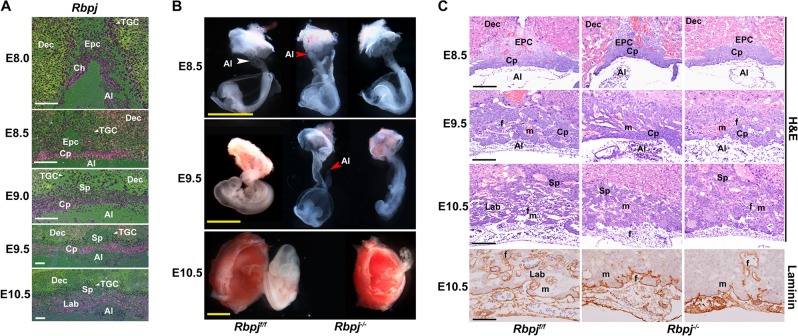
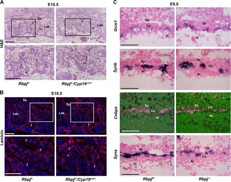
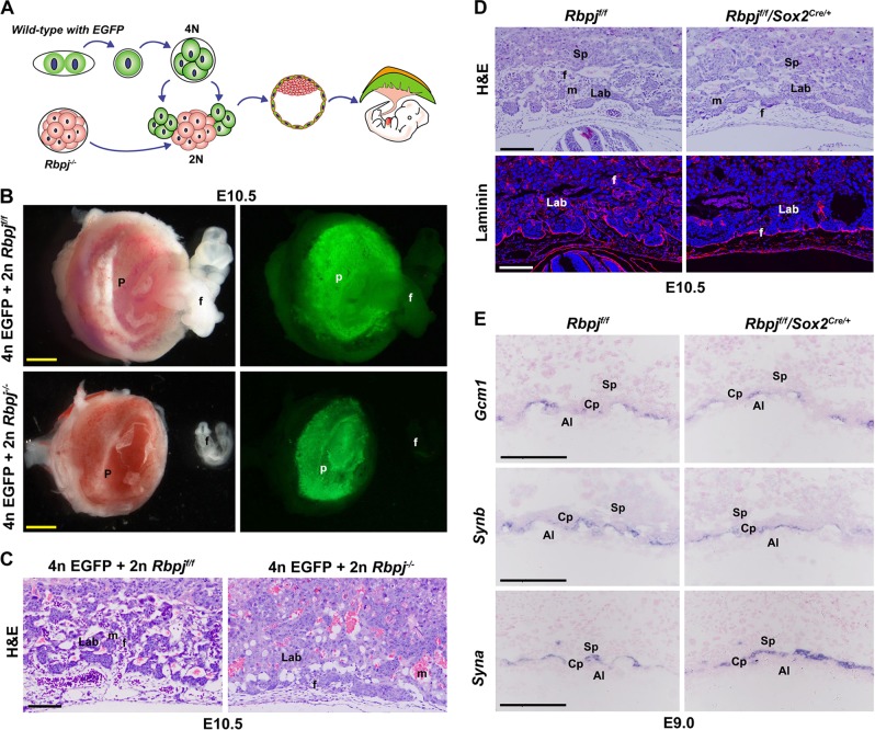
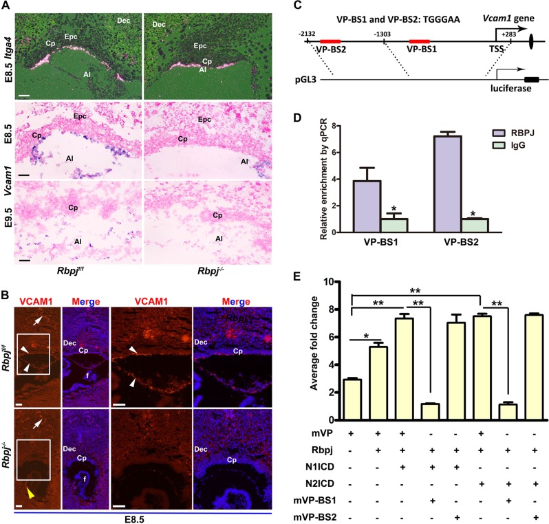
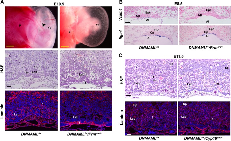
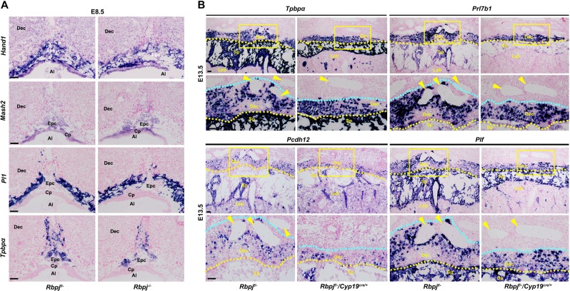
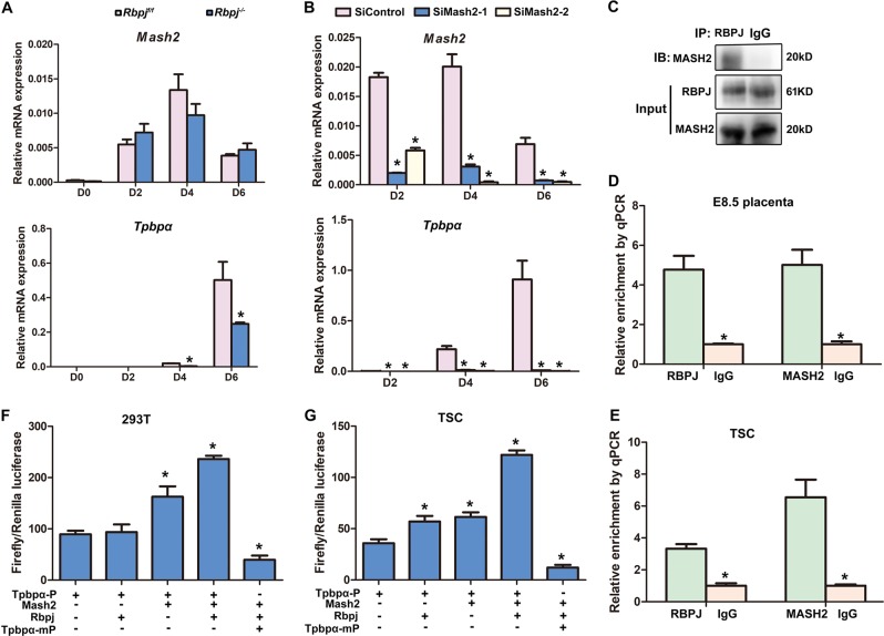
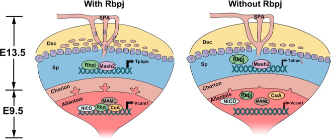
The placenta, responsible for the nutrient and gas exchange between the mother and fetus, is pivotal for successful pregnancy. It has been shown that Rbpj, the core transcriptional mediator of Notch signaling pathway, is required for normal placentation in mice. However, it remains largely unclear how Rbpj signaling in different placental compartments coordinates with other important regulators to ensure normal placental morphogenesis. In this study, we found that systemic deletion of Rbpj led to abnormal chorioallantoic morphogenesis and defective trophoblast differentiation in the ectoplacental cone (EPC). Employing mouse models with selective deletion of Rbpj in the allantois versus trophoblast, combining tetraploid aggregation assay, we demonstrated that allantois-expressed Rbpj is essential for chorioallantoic attachment and subsequent invagination of allantoic blood vessels into the chorionic ectoderm. Further studies uncovered that allantoic Rbpj regulates chorioallantoic fusion and morphogenesis via targeting Vcam1 in a Notch-dependent manner. Meanwhile, we also revealed that trophoblast-expressed Rbpj in EPC facilitates Mash2's transcriptional activity, promoting the specification of Tpbpα-positive trophoblasts, which differentiate into trophoblast subtypes responsible for interstitial and endovascular invasion at the later stage of placental development. Collectively, our study further shed light on the molecular network governing placental development and functions, highlighting the necessity of a spatiotemporal coordination of Rbpj signaling for normal placental morphogenesis.


Conflict of interest statement

The authors declare that they have no conflict of interest.

Figures

Fig. 1
Fig. 1. Rbpj deficiency derails normal placental development.
a In situ hybridization analysis of Rbpj mRNA expression during early placental development from E8.0 to E10.5. White arrowheads indicate TGCs. Images are representative of two independent experiments. b Morphological appearance of the conceptus dissected and removed from the parietal yolk sac at E8.5, and partially dissected feto-placental units at E9.5 and E10.5. The white arrowhead indicates the fused allantois, while the red arrowheads indicate the ends of the allantois failing to fuse with the chorion and appearing as a bud in Rbpj−/− mutants. c Histological sections of Rbpjf/f and Rbpj−/− chorionic plates and allantois with hematoxylin–eosin (HE) and Laminin staining from E8.5 to E10.5. The fetal vessels were indicated by Laminin staining. Images in b and c are representative of at least three independent experiments. Al allantois, Ch chorion, Cp chorion plate, Epc ectoplacental cone, Dec decidual basalis, TGC trophoblast giant cell, Lab labyrinth, Sp spongiotrophoblast layer, f fetal blood vessel, m maternal blood sinus. Yellow scale bars: 1 mm; white and black scale bars: 100 μm
Fig. 2
Fig. 2. Trophoblast-expressed Rbpj is dispensable for chorioallantoic fusion and branching morphogenesis.
HE (a) and Laminin staining (b) of E10.5 placenta with Rbpj deleted specifically in placental trophoblast cells by Cyp19cre/+ transgenic mice model. In order to achieve efficient Rbpj deletion in trophoblast cells, we first deleted one allele of Rbpj systemically before deleting the other allele by Cyp19cre/+. Cy3-labeled laminin is in red, DAPI-labeled nuclei in blue. Regions of interest are boxed in black (a) or white (b), and magnified below. c The differentiation of the chorionic trophoblast was revealed by the expressions of Gcm1, Synb, and Cebpα (markers for SynT-II), and Syna (marker for SynT-I) in Rbpjf/f and Rbpj−/− chorion plate (Cp). The expression of these markers was comparable between the control (Rbpjf/f) and Rbpj-null mutants in in situ hybridization assay. Images in ac are representative of at least three independent experiments. Al allantois, Cp chorion plate, Lab labyrinth, Sp spongiotrophoblast layer; f fetal blood vessel; m, maternal blood sinus. White and black scale bars: 100 μm
Fig. 3
Fig. 3. Allantois-expressed Rbpj is essential for chorioallantoic fusion and branching morphogenesis.
a Diagram illustrating the procedure of tetraploid aggregation, in which one Rbpj−/− morula aggregated with two wild-type tetraploid EGFP-expressing four-cell embryos. The tetraploid cells contribute exclusively to the trophoblast cells of the placenta, rather than the allantois. b E10.5 embryos and placentas generated by tetraploid aggregation assay. c HE staining of E10.5 reconstructed placentas from tetraploid aggregation assay. d Shallow invagination of allantoic blood vessels into the chorionic ectoderm was revealed by HE and laminin staining in placentas with allantois-specific deletion of Rbpj by Sox2cre/+. Cy3-labeled Laminin is in red, DAPI-labeled nuclei in blue. e The differentiation of a chorionic trophoblast was revealed by the expressions of Gcm1 and Synb (markers for SynT-II), and Syna (marker for SynT-I). By in situ hybridization, the expression of these markers was comparable between the Rbpjf/f and Rbpjf/f/Sox2cre/+ placentas. Images in b–e are representative of at least three independent experiments. Al allantois, Cp chorion plate, Lab labyrinth, f fetal blood vessel, m maternal blood sinus, p placenta, Sp spongiotrophoblast layer. Yellow scale bars: 1 mm; white and black scale bars: 100 μm
Fig. 4
Fig. 4. Rbpj directs allantoic Vcam1 expression in a Notch-dependent manner.
a The expression of Itga4 and Vcam1 was detected by in situ hybridization in the controls (Rbpjf/f) versus Rbpj−/− mutants. b VCAM1 expression at protein level was revealed in E8.5 control (Rbpjf/f) and Rbpj−/− chorion plate. Cy3-labeled VCAM1 is in red, DAPI-labeled nuclei in blue. White arrows indicate VCAM1 expression in maternal decidual vascular endothelial cells. White arrowheads, the VCAM1 expression in allantoic cells. Yellow arrowheads, the VCAM1 expression in the fetus. Regions of interest are boxed in white and magnified on the right. c Schematic representation of the promoter region of Vcam1 and PGL3-Vcam1 constructs. The red rectangle indicated two potential Rbpj-binding sites within a region spanning 2 kb upstream of the TSS of Vcam1. d ChIP assay revealed that Rbpj was enriched at the promoter region of Vcam1 in E8.5 placentas. N = 3. e The activity of the Vcam1 promoter was activated by overexpression of Rbpj in combination with NICD (NICD1 or NICD2) overexpression or not. Mutation analysis revealed that only VP-BS1 responded to the activation of Rbpj-mediated Notch signaling. N = 3. *P < 0.05, **P < 0.01. Images in a and b are representative of at least three independent experiments. Al allantois, Cp chorion plate, Dec decidual basalis, Epc ectoplacental cone, f fetus. Scale bars: 100 μm
Fig. 5
Fig. 5. Genetic evidence for Rbpj-mediated Notch signaling pathway regulating chorioallantoic morphogenesis.
a Whole-mount views, as well as HE and Laminin staining of DNMAMLf/+ and DNMAMLf/+/Prmcre/+ placentas and yolk sacs. Note that the expression of dominant negative MAML (DNMAML), which blocks Notch-Rbpj signaling, disturbs normal chorioallantoic branching morphogenesis. Black arrowheads indicated the large vitelline vessels in the yolk sac. Cy3-labeled Laminin is in red, DAPI-labeled nuclei in blue. b In situ hybridization analysis of Itga4 and Vcam1 expression. Note the disappeared expression of Vcam1 in the allantois of DNMAMLf/+/Prmcre/+ placentas. c HE and laminin staining of DNMAMLf/+ and DNMAMLf/+/Cyp19cre/+ placentas. Note the well-intermingled maternal sinuses and the invading fetal vessels filled with nucleated erythrocytes in E10.5 DNMAMLf/+/Cyp19cre/+ placentas. Cy3-labeled Laminin is in red, DAPI-labeled nuclei in blue. Images in ac are representative of at least three independent experiments. Al allantois, Cp chorion plate, Lab labyrinth, Sp spongiotrophoblast, f fetal blood vessel, m maternal blood sinus, p placenta, Ys yolk sac. Yellow scale bars: 1 mm; white and black scale bars: 100 μm
Fig. 6
Fig. 6. Rbpj deficiency attenuates the specification of Tpbpα-positive trophoblast.
a The expression of trophoblast marker genes was detected by in situ hybridization analysis in the control (Rbpjf/−) versus Rbpj-null ectoplacental core at E8.5. The Hand1 expression was unchanged by Rbpj deletion, correlating with normal differentiation of Pl1-expressing giant cells. While the expression of Mash2 was unaffected by Rbpj deficiency, the Tpbpα-positive spongiotrophoblast cells were decreased significantly. b In situ hybridization analysis revealed that the invasive ability of trophoblast cells differentiated from the Tpbpα-positive trophoblast cells in the ectoplacental core was disturbed by Rbpj deletion. Prl7b1 marks both the invasive glycogen trophoblast cells and SPA-TGCs. Pcdh12 marks glycogen trophoblast cells and Plf marks SPA-TGCs. Yellow arrowheads indicate the maternal spiral arteries. Yellow dotted lines indicate the interface between the maternal decidua and the spongiotrophoblast layer, while the light blue dotted lines show the boundaries that the trophoblast invades to the maternal decidua. The distance of trophoblast invasion is between the yellow and light blue lines. Regions of interest are boxed in yellow and magnified below. Images in a and b are representative of at least three independent experiments. Al allantois, Cp chorion plate, Epc ectoplacental cone, Dec decidual basalis, Lab labyrinth, Sp spongiotrophoblast. Scale bars: 100 μm
Fig. 7
Fig. 7. Rbpj facilitates Mash2 to promote the specification of Tpbpα-positive trophoblast.
a Quantitative RT-PCR analysis of Mash2 and Tpbpα during the differentiation of Rbpjf/f and Rbpj−/− trophoblast stem cells. b The expression of Mash2 and Tpbpα after Mash2 knockdown via siRNA was detected by quantitative RT-PCR, during trophoblast stem cell differentiation. Values are normalized by GAPDH expression level and indicated as mean ± SEM. N = 3. *P < 0.05. c The interactions between the Rbpj and Mash2 were revealed by IP assay. d, e Rbpj and Mash2 were enriched at the promoter region of Tpbpα in both the E8.5 placentas (d) and in vitro-cultured trophoblast cells (e). f, g Rbpj facilitates Mash2 to activate the promoter activity of Tpbpα in both 293 T cells (f) and trophoblast stem cells (g), while deletion of Mash2 binding site in Tpbpα regulatory region diminished the activity of Tpbpα promoter increased by Rbpj and Mash2. *P < 0.05. Data in ag are representative of at least three independent experiments
Fig. 8
Fig. 8. Diagram illustrating the functions of RBPJ-mediated signaling during chorioallantoic development and trophoblast differentiation in EPC (Sp).
In the presence of Rbpj (with Rbpj), Rbpj regulates the expression of Vcam1 in the allantois through a Notch-dependent manner to ensure normal chorioallantoic branching morphogenesis, and facilitated Mash2 in EPC (Sp) to promote the specification of Tpbpα-positive trophoblast cells simultaneously. However, failed chorioallantoic branching and defective trophoblast differentiation in EPC (Sp) were observed in the absence of Rbpj (without Rbpj). Dec decidual basalis, Sp spongiotrophoblast layer, SPA spiral arteries

References

    1. Rossant J, Cross JC. Placental development: lessons from mouse mutants. Nat. Rev. Genet. 2001;2:538–548. doi: 10.1038/35080570. - DOI - PubMed
    1. Watson ED, Cross JC. Development of structures and transport functions in the mouse placenta. Physiology. 2005;20:180–193. doi: 10.1152/physiol.00001.2005. - DOI - PubMed
    1. Maltepe E, Bakardjiev AI, Fisher SJ. The placenta: transcriptional, epigenetic, and physiological integration during development. J. Clin. Invest. 2010;120:1016–1025. doi: 10.1172/JCI41211. - DOI - PMC - PubMed
    1. Rai A, Cross JC. Development of the hemochorial maternal vascular spaces in the placenta through endothelial and vasculogenic mimicry. Dev. Biol. 2014;387:131–141. doi: 10.1016/j.ydbio.2014.01.015. - DOI - PubMed
    1. Downs KM. Early placental ontogeny in the mouse. Placenta. 2002;23:116–131. doi: 10.1053/plac.2001.0763. - DOI - PubMed

Publication types

MeSH terms

Substances