. 2019 Jun 5;9(1):8307.
doi: 10.1038/s41598-019-44407-9.

Protein kinase A inhibits tumor mutator APOBEC3B through phosphorylation

Affiliations

Protein kinase A inhibits tumor mutator APOBEC3B through phosphorylation

Tadahiko Matsumoto et al. Sci Rep. .

Abstract

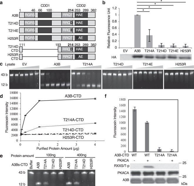
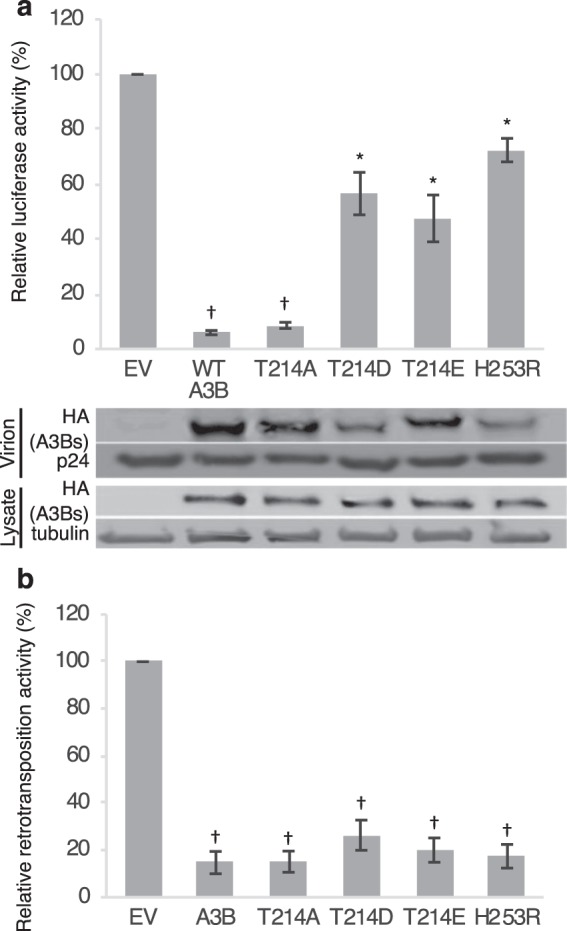
APOBEC3B cytidine deaminase (A3B) catalyzes cytosine into uracil in single-strand DNA and induces C-to-T mutations in genomic DNA of various types of tumors. Accumulation of APOBEC signature mutations is correlated with a worse prognosis for patients with breast cancer or multiple myeloma, suggesting that A3B activity might be a cause of the unfavorable DNA mutations and clonal evolution in these tumors. Phosphorylation of conserved threonine residues of other cytidine deaminases, activation induced deaminase (AID) and APOBEC3G, inhibits their activity. Here we show that protein kinase A (PKA) physically binds to A3B and phosphorylates Thr214. In vitro deaminase assays and foreign DNA editing assays in cells confirm that phosphomimetic A3B mutants, T214D and T214E, completely lose deaminase activity. Molecular dynamics simulation of A3B phosphorylation reveals that Thr214 phosphorylation disrupts binding between the phospho-A3B catalytic core and ssDNA. These mutants still inhibit retroviral infectivity at least partially, and also retain full anti-retrotransposition activity. These results imply that PKA-mediated phosphorylation inhibits A3B mutagenic activity without destructing its innate immune functions. Therefore, PKA activation could reduce further accumulation of mutations in A3B overexpressing tumors.


Conflict of interest statement

The authors declare no competing interests.

Figures

Figure 1
Figure 1
PKACA phosphorylates A3B Thr214 in vivo and in vitro. (a) Schematic figure of AID, A3G, A3B and their mutants. A3B WT CTD and A3B T214A CTD are the purified protein constructs. (b,c) A3B physically binds to PKACA. HEK293T cells were transfected with expression vectors for A3B-HA and FLAG-PKACA. Lysates from these cells were co-immunoprecipitated with anti-HA (b) or anti-FLAG antibodies (c), followed by immunoblot with the indicated antibodies. (d) PKACA phosphorylates A3B in HEK293T cells. HEK293T cells were transfected with expression vectors for A3B, A3G, and A3G DM with or without PKACA, as shown above. 36hrs after transfection, we semi-purified the APOBECs by immunoprecipitation, and PKA-mediated phosphorylation was detected by immunoblot with anti-RXXS/T-p antibodies. (e,f) PKACA phosphorylates Thr214 of A3B. (e) Expression vectors for A3B alanine mutants with or without PKACA are introduced in HEK293T cells as indicated. 36 hrs after transfection, cells were collected, lysed, immunoprecipitated with the anti-HA antibody and subjected to immunoblot with the indicated antibodies. The results show that A3B Thr214 is a crucial residue for PKA-mediated phosphorylation. (f) In vitro phosphorylation assays show that PKACA directly phosphorylates A3B-CTD, but not A3B T214A CTD. AID, activation-induced deaminase; WT, wild type; DM, double mutant; IP, immunoprecipitation.
Figure 2
Figure 2
Phosphomimetic A3B mutants lose their CDA in vitro. (a) Schematic figure of phosphomimetic A3B mutants at Thr214. T214D CTD and H253R CTD are the purified protein constructs. (b) Phosphomimetic A3B mutants lose their CDA in vitro. FRET-based CDA assays were performed using lysates from HEK293T cells with exogenously-expressed A3B or its mutants. Each of the fluorescent intensity numerical values represent the average of three independent experiments, normalized to that of the WT sample. Bars indicate standard error (SE), and asterisks (*) show statistically significant difference (p < 0.05) (upper panel). The protein levels of the A3B mutants used in the experiment were comparable (lower panel). (c) Phosphomimetic A3B mutants lose CDA against the TCA sequence in the gel-based CDA assay. In this assay A3B CDA catalyzes substrate 43b oligo to 12b oligo transition. Lysates from cells transfected with EV, T214D, T214E or H253R have no CDA, and those with T214A have less CDA than those with WT. (d) FRET-based CDA assays using purified proteins. Purified C-terminal A3B T214D almost completely loses CDA in vitro. Each of the fluorescent intensity numerical values represent the average of three independent experiments. (e) Gel-based CDA assay using purified proteins. A3B T214D loses CDA, and T214A has weak CDA against the TCA sequence. (f) Phosphorylation of A3B reduces its cytidine deaminase activity. A3B-CTD and T214A-CTD were phosphorylated by PKACA in vitro, and subsequently performed in vitro CDA assays. Values represent the average of two independent experiments, and bars indicate SE.
Figure 3
Figure 3
A3B phosphorylation or mutation affects ssDNA binding. Structural models for A3Bs in an ssDNA-bound state were constructed with reported X-ray crystal structures of A3B (PDB number 5CQH) and ssDNA (PDB number 5TD5), and subjected to MDS as described in the experimental procedures section. (a) A structure in an ssDNA-bound state at thermodynamic equilibrium, for 80 ns of MDS of the A3B WT-ssDNA complex model. The active site of the enzyme is highlighted. A3B residues (T214, H253, E255, C284, and C289), located near cytosine and Zn, and presumably involved in the chemical catalysis of DNA cytidine deamination, are shown in color sticks. Orange dotted lines indicate hydrogen bonds between Thr214 and cytosine. (b) Binding modes of ssDNA with various A3Bs (A3B WT, phosphorylated, T214D, T214E, T214A, and H253R) after 80 ns of MDS. A3B active centers around Zn and ssDNA are highlighted.
Figure 4
Figure 4
Phosphomimetic A3B mutants lose foreign DNA editing activity. (a) 3D-PCR of EGFP exposed to WT A3B and its mutants. HEK293T cells were co-transfected with expression vectors for A3B or its mutants together with pDON-EGFP and pEF-UGI. Total DNA was recovered 2 days after transfection, and the EGFP gene was amplified by 3D-PCR at the indicated denaturation temperatures (Td). (b) Expression levels of A3B WT and its mutants were comparable. (c) Mutational matrix of EGFP sequences derived from PCR products amplified at the lowest Td in each sample. (d) Dinucleotide context of foreign DNA editing. The rates of C-to-T transitions in the indicated dinucleotide sequence are shown.
Figure 5
Figure 5
Phosphomimetic A3B mutants retain anti-retroviral activity and retrotransposition restriction. (a) VSV-G pseudotyped NL4-3 viruses were prepared by co-transfection of pNL4-3/Δenv/luc and VSV-G together with expression vectors for A3B or its mutants. HEK293T cells were infected with the produced viruses. Luciferase activity of the target cell lysate was measured and presented as ratio of infectivity reported to the EV (upper panel). Protein levels of A3B and mutants within the lysates or virions are comparable (lower panel) (b) A3B suppresses retrotransposition. Both WT and phosphomimetic mutants, and even the catalytically inactive H253R mutant, inhibit retrotransposition. Values represent the average of three independent experiments, bars indicate SE, and daggers (†) and asterisks (*) show statistically significant difference (p < 0.01 and p < 0.05, respectively).

Similar articles

Cited by

References

    1. Harris RS, Hultquist JF, Evans DT. The restriction factors of human immunodeficiency virus. J Biol Chem. 2012;287:40875–40883. doi: 10.1074/jbc.R112.416925. - DOI - PMC - PubMed
    1. Bishop KN, et al. Cytidine deamination of retroviral DNA by diverse APOBEC proteins. Curr Biol. 2004;14:1392–1396. doi: 10.1016/j.cub.2004.06.057. - DOI - PubMed
    1. Bogerd HP, Wiegand HL, Doehle BP, Cullen BR. The intrinsic antiretroviral factor APOBEC3B contains two enzymatically active cytidine deaminase domains. Virology. 2007;364:486–493. doi: 10.1016/j.virol.2007.03.019. - DOI - PMC - PubMed
    1. Xu Z, Zan H, Pone EJ, Mai T, Casali P. Immunoglobulin class-switch DNA recombination: induction, targeting and beyond. Nat Rev Immunol. 2012;12:517–531. doi: 10.1038/nri3216. - DOI - PMC - PubMed
    1. Harris RS, et al. DNA Deamination Mediates Innate Immunity to Retroviral Infection. Cell. 2003;113:803–809. doi: 10.1016/s0092-8674(03)00423-9. - DOI - PubMed

Publication types