. 2019 Aug;17(8):513-525.
doi: 10.1038/s41579-019-0204-7.

CRISPR-Cas in mobile genetic elements: counter-defence and beyond

Affiliations

CRISPR-Cas in mobile genetic elements: counter-defence and beyond

Guilhem Faure et al. Nat Rev Microbiol. 2019 Aug.

Abstract

The principal function of CRISPR-Cas systems in archaea and bacteria is defence against mobile genetic elements (MGEs), including viruses, plasmids and transposons. However, the relationships between CRISPR-Cas and MGEs are far more complex. Several classes of MGE contributed to the origin and evolution of CRISPR-Cas, and, conversely, CRISPR-Cas systems and their components were recruited by various MGEs for functions that remain largely uncharacterized. In this Analysis article, we investigate and substantially expand the range of CRISPR-Cas components carried by MGEs. Three groups of Tn7-like transposable elements encode 'minimal' type I CRISPR-Cas derivatives capable of target recognition but not cleavage, and another group encodes an inactivated type V variant. These partially inactivated CRISPR-Cas variants might mediate guide RNA-dependent integration of the respective transposons. Numerous plasmids and some prophages encode type IV systems, with similar predicted properties, that appear to contribute to competition among plasmids and between plasmids and viruses. Many prokaryotic viruses also carry CRISPR mini-arrays, some of which recognize other viruses and are implicated in inter-virus conflicts, and solitary repeat units, which could inhibit host CRISPR-Cas systems.


Figures

Fig. 1 ∣
Fig. 1 ∣. Evolutionary relationships and gene fluxes between CRISPR–Cas systems and mobile genetic elements.
The evolutionary reconstruction of the fluxes of genes from mobile genetic elements (MGEs) to class 1 and class 2 CRISPR–Cas systems (In) and from CRISPR–Cas systems to MGEs (Out) is shown. The gene flux directions are indicated by vertical black arrows. The types of donor and recipient MGE are indicated on the left, and the characteristic gene architecture is schematically shown for each type. iscB and tnpB are RuvC-like nuclease domain-containing proteins encoded by non-autonomous transposable elements (also known as insertion sequences (IS)) that appear to be the ancestors of cas9 and cas12, respectively. The gene repertoires of Class 1 and Class 2 CRISPR–Cas systems are schematically shown between the ‘In’ and ‘Out’ panels. Genes are displayed as block arrows. Homologous genes are colour-coded and identified by the corresponding protein family names, except for the effector module genes. The cas gene names follow the classification from REF.. tnsB and tnsC are the genes from Tn7-like transposons. Genes other than cas and tns are shown by grey block arrows. CRISPR arrays are shown as grey rectangles (repeats) and coloured diamonds (spacers). The schemes include the union of the key components of the different CRISPR–Cas types in each of the classes that do not necessarily co-occur within individual CRISPR–cas loci (for example, tnsB and tnsC that represent a putative alternative adaptation module do not co-occur with cas1, cas2 and cas4). In the ‘Out’ panel, subtypes of the CRISPR–Cas systems acquired by MGEs are indicated below the schematic representations of the gene architectures of the respective MGE. HEPN, RNase of the Higher Eukaryotes and Prokaryotes Nucleotide-binding superfamily; LE, RE, cis-acting left and right terminal sequences of Tn7-like transposons; RT, reverse transcriptase; TR, terminal repeats.
Fig. 2 ∣
Fig. 2 ∣. Derived type I CRISPR–Cas systems in Tn7-like transposons.
Genomic architectures of Tn7-like transposon loci encoding derived type I CRISPR–Cas systems are compared with typical systems of the same type. a ∣ Architectures of the canonical subtype I-F CRISPR–cas locus and the derived, Tn7-encoded minimal I-F variant. The transposon-encoded variant lacks the cas3 gene and thus is not competent for interference, and is hypothesized to instead facilitate integration in a CRISPR RNA (crRNA)-dependent manner. b ∣ Architectures of the canonical subtype I-B CRISPR–cas locus and the derived, Tn7-encoded I-B variant. Similar to the independently evolved, ‘minimal’ subtype I-F variant, transposon-encoded I-B variants lack the cas3 gene and thus are not competent for interference, and are hypothesized instead to facilitate integration in a crRNA-dependent manner. c ∣ Architecture of subtype V-U5 CRISPR–Cas systems in cyanobacterial Tn7-like transposons. The V-U5 effectors are predicted to be inactive due to replacement of the catalytic amino acid residues in the RuvC-like nuclease domain. Thus, these transposon-encoded systems are predicted to be incapable of interference but might facilitate integration in a crRNA-dependent manner, similar to the subtype I-F and I-B variants. d ∣ Architectures of the canonical subtype I-E CRISPR–cas locus and the derived subtype I-E variant in Streptomycetaceae associated with the tnsBC genes. The TnsB and TnsC proteins might comprise an alternative CRISPR adaptation module. Genes are displayed as block arrows. Homologous genes are colour-coded and are identified by family names. Light grey arrows denote representative uncharacterized genes, but additional genes not conserved in these regions are omitted for clarity. The cas gene names follow the classification from REF.. HD is the effector nuclease domain that belongs to the HD family of nucleases (named after the respective dyad of catalytic amino acid residues). CRISPR arrays are shown as grey rectangles (repeats) and coloured diamonds (spacers).The products of the tnsA, tnsB, tnsC and tniQ(tnsD) transposon genes are responsible for the transposition of Tn7-like elements. LE and RE (brown rectangles) denote the cis-acting left and right terminal sequences of Tn7-like transposons, respectively. The tniQ(tnsD)-encoded proteins function with other tns gene products to direct transposon insertion into att (attachment or integration) sites in the host genome that are specific for each Tn7-like transposon subtype.
Fig. 3 ∣
Fig. 3 ∣. Type IV CRISPR–Cas systems in plasmids and prophages.
Genomic architectures of type IV CRISPR–cas Loci are shown. Genes and CRISPR arrays are shown as in FIG. 1. a ∣ Architectures of the two subtypes of type IV CRISPR–Cas systems. b ∣ Type IV CRISPR–cas Loci in two prophages and a plasmid containing cysH genes (yeLLow). Species, accession numbers and coordinates are indicated to the right of each Locus. c ∣ Prevalence of the cysH gene in subtypes IV-A and IV-B. The coLours represent the presence (bLue) and absence (orange) of the cysH gene within the subtype IV CRISPR–cas Loci. SS, smaLL subunit of the effector compLex.
Fig. 4 ∣
Fig. 4 ∣. Origins of spacers in CRISPR arrays from viruses and proviruses.
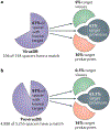
a ∣ CRISPR spacers with matching protospacers identified in the virus database (VirusDB). VirusDB is compiled from all prokaryotic virus sequences extracted from the National Center for Biotechnology Information (NCBI) nucleotide database in August 2018 (see Supplementary Dataset 5 for details), b ∣ CRISPR spacers with matching protospacers identified from the provirus database (ProvirusDB). ProvirusDB consists of provirus sequences predicted within prokaryotic genomes that are available in the NCBI nucleotide sequence database (see Supplementary Methods for details). The pie charts on the left show the fractions of spacers for which protospacers were identified. The diagrams on the right show the breakdown of the identified protospacers between prokaryotes (pink), proviruses (grey) and viruses (green).
Fig. 5 ∣
Fig. 5 ∣. CRISPR mini-arrays in Streptococcus thermophilus lytic bacteriophages.
An example of a CRISPR mini-array that is conserved in a group of Streptococcus thermophilus bacteriophages and contains spacers cross-targeting viruses of the same group but avoiding self-targeting is depicted. Genes are shown by yellow block arrows. The predicted promoters of the CRISPR arrays are shown by thick red lines in the viruses and by a thick grey line in the host, the repeats are shown as red vertical rectangles and the spacers are shown as coloured diamonds. Distal repeats (partial repeats located at the 3′ ends of the mini-arrays) are shown as pink or brown rectangles (for the phage mini-arrays and the host array, respectively). A small coloured rectangle in the 3′ portion of the partial repeat shows similar (between phages, light grey) or distinct (light grey in phages and dark grey in the host) distal partial repeat sequences. The isolated red vertical rectangle indicates the anti-repeat region of the transactivating crRNA (tracrRNA) in the host CRISPR–cas locus. The protospacers in phage genes are shown by vertical rectangles in genes indicating the identified protospacers that match the colours of the respective spacers. a ∣ The CRISPR mini-array loci in S. thermophilus phages are shown at the top, and the host CRISPR–cas locus is shown underneath.The phage-encoded CRISPR mini-arrays are shown inside the black rectangular outline and are located between the genes encoding the small (TerS) and large (TerL) subunits of the terminase in five closely related phages (names indicated). The dashed arrows indicate the cross-targeting of phage genes by spacers from mini-arrays of other phages (the names of four targeted phages are indicated for each arrow; in cases when more than four phages were targeted, four representatives were selected randomly). The grey arrows show the hypothesized (indicated by question marks) recruitment of host Cas9 and tracrRNA for processing of the phage crRNAs and interference between phages. b ∣ TerL-targeting spacers from phage mini-arrays with a perfect match to a related phage but a partial match to the mini-array-carrying phage (blue protospacer in part a). Mismatching nucleotides are highlighted. The arrows indicate cross-targeting.
Fig. 6 ∣
Fig. 6 ∣. Proposed functions and mechanisms of virus-encoded CRISPR mini-arrays and solitary repeat units.
Three distinct functions are proposed for CRISPR mini-arrays and solitary repeat units (SRUs). Viruses are denoted by a schematic image of a tailed phage that is shown attached to the cell wall of a bacterium or archaeon. Viral DNA that is injected into the host cell is shown by a curved descending line. In the CRISPR arrays (virus mini-array and host array), repeats are shown as vertical red rectangles, and the distal, degraded repeat is shown by a smaller rectangle (pink for viruses and dark brown for the host); spacers are shown by coloured diamonds. The spacer sequences in the (pre-)CRISPR RNA ((pre-)crRNA) are shown in blue, and repeats are shown in red. The host interference module is shown in yellow, with genes denoted by block arrows and the effector proteins shown as lobe shapes, and the genes of the adaptation module are shown by grey block arrows. Right-angled arrows indicate transcription. In the crRNA and pre-crRNA, spacers are shown in blue, repeats are shown in red and the partial repeats are shown in pink. a ∣ Inter-virus competition. Spacers from virus mini-arrays target genomes of related viruses and thus prevent their reproduction in the same cell. In this model, virus and provirus mini-arrays with repeats (nearly) identical to those in the host arrays are compatible with the host interference machinery. Virus or provirus crRNA is loaded onto the host effector complex (protein) to target the DNA of superinfecting viruses containing the matching protospacer sequences, thus, providing immunity against these viruses. In the case of lytic viruses (part Aa), there is a transient symbiosis between the host and the mini-array-carrying virus, resulting in restriction of superinfection by competing viruses, such that all of the resources of the host are appropriated by the mini-array-carrying virus that eventually kills the host. In the case of proviruses (part Ab), there is a long-term symbiosis whereby the provirus-encoded CRISPR mini-array prevents superinfection by competing viruses. The provirus is shown in brackets. B ∣ Inhibition of the host CRISPR–Cas system by a virus-encoded SRU. In this model, expression of a virus-encoded SRU generates a partial crRNA that lacks a spacer but binds to the host interference complex and acts as a dominant negative inhibitor of the host CRISPR–Cas. C ∣ Integration of a virus genome into the host CRISPR array via homologous recombination with an SRU or mini-array. In this model, a viral SRU or mini-array recombines with the identical repeat in the host CRISPR array, resulting in integration of the provirus (shown in brackets) inside the CRISPR array. The provirus would abrogate the transcription of the portion of the CRISPR array downstream of the integration point and inactivate host immunity.

References

    1. Makarova KS et al. An updated evolutionary classification of CRISPR–Cas systems. Nat. Rev. Microbiol 13, 722–736 (2015). - PMC - PubMed
    1. Mohanraju P. et al. Diverse evolutionary roots and mechanistic variations of the CRISPR-Cas systems. Science 353, aad5147 (2016). - PubMed
    1. Barrangou R & Horvath P A decade of discovery: CRISPR functions and applications. Nat. Microbiol 2, 17092 (2017). - PubMed
    1. Jackson SA et al. CRISPR-Cas: adapting to change. Science 356, eaal5056 (2017). - PubMed
    1. Koonin EV, Makarova KS & Zhang F Diversity, classification and evolution of CRISPR-Cas systems. Curr. Opin. Microbiol 37, 67–78 (2017).

      This article is the latest published overview of the CRISPR–Cas diversity, with an emphasis on Class 2 systems discovered through dedicated search efforts.

Publication types

Substances