. 2019 Aug 1;317(2):F303-F321.
doi: 10.1152/ajprenal.00214.2019. Epub 2019 Jun 5.

Expression and distribution of PIEZO1 in the mouse urinary tract

Affiliations

Expression and distribution of PIEZO1 in the mouse urinary tract

Marianela G Dalghi et al. Am J Physiol Renal Physiol. .

Abstract

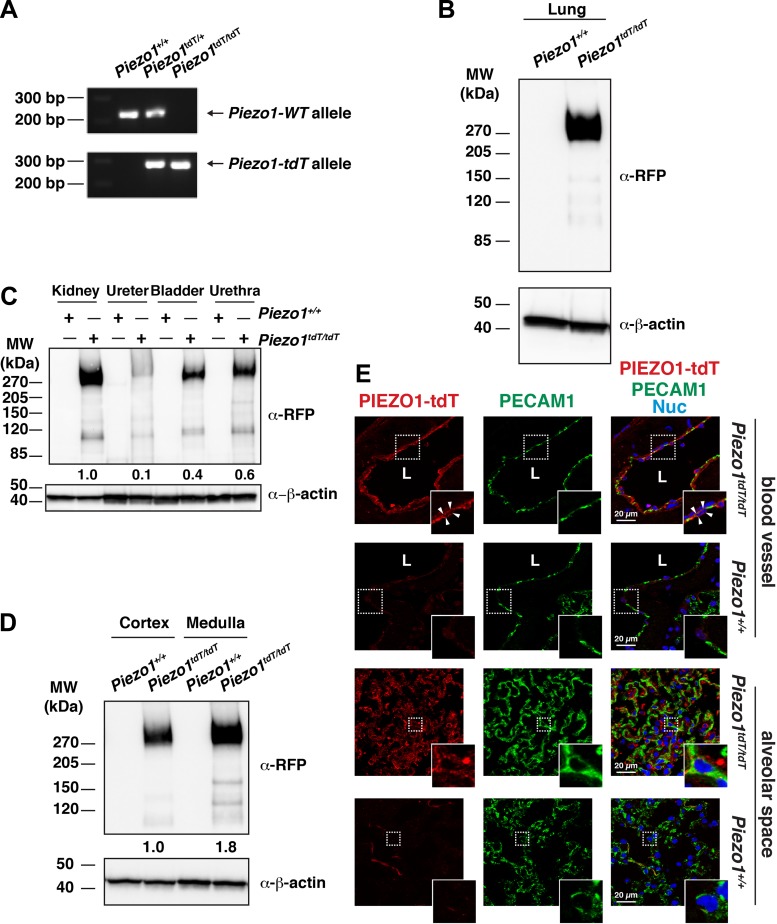
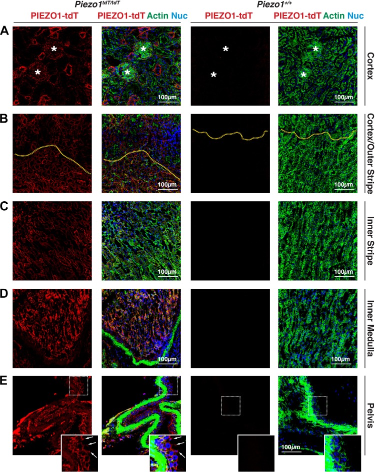
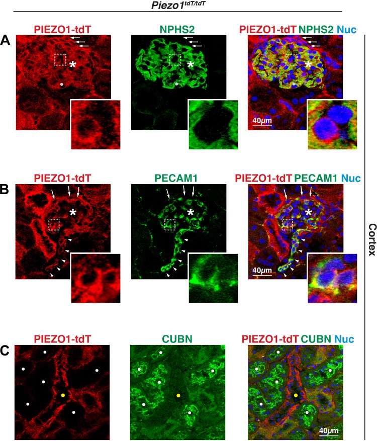
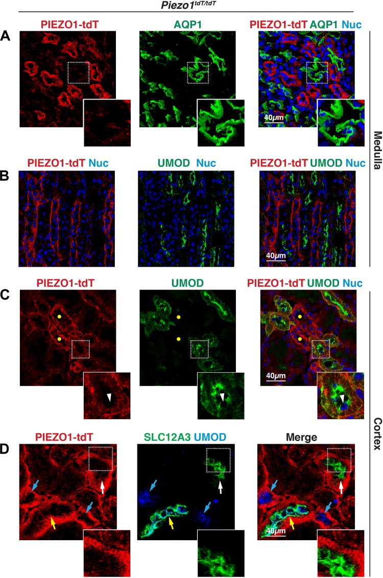
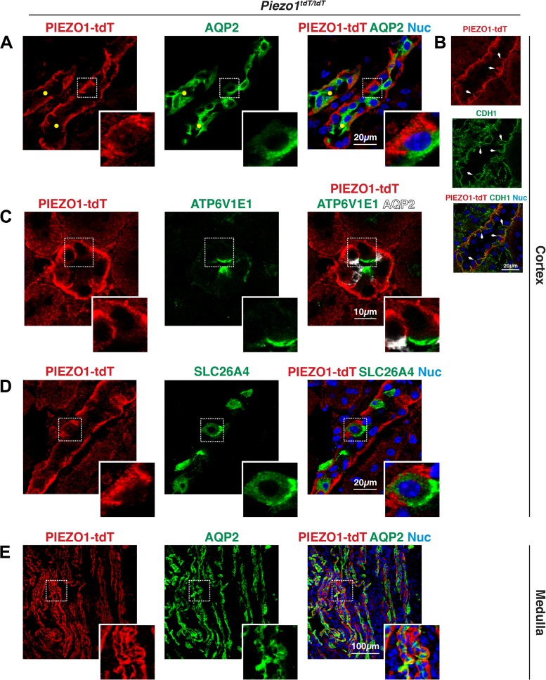
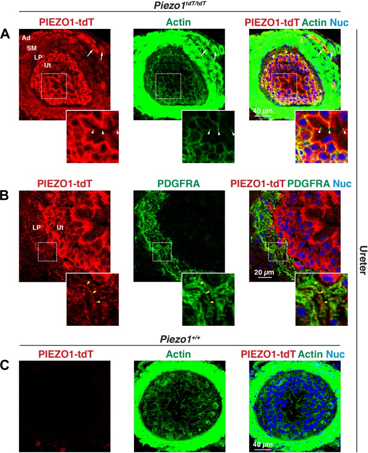
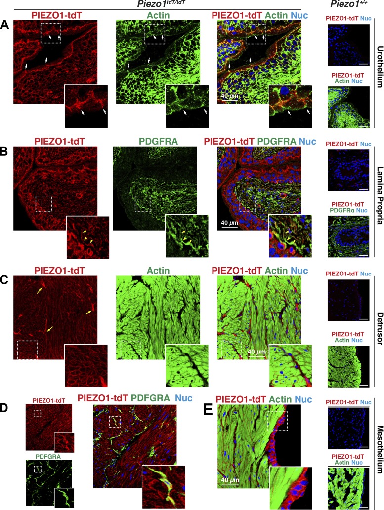
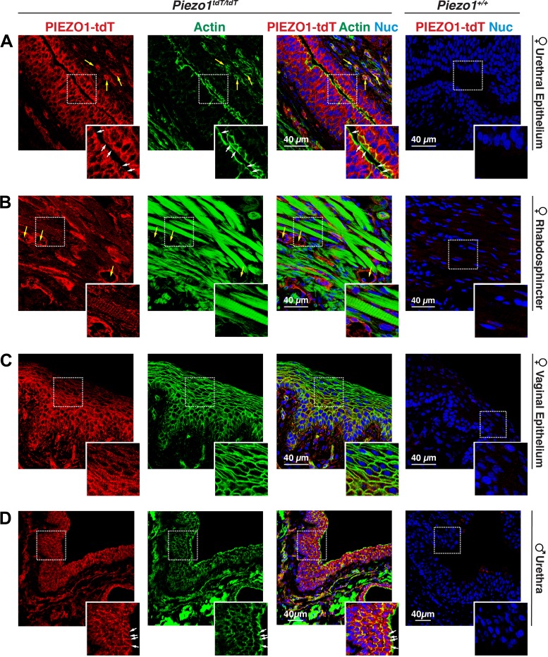
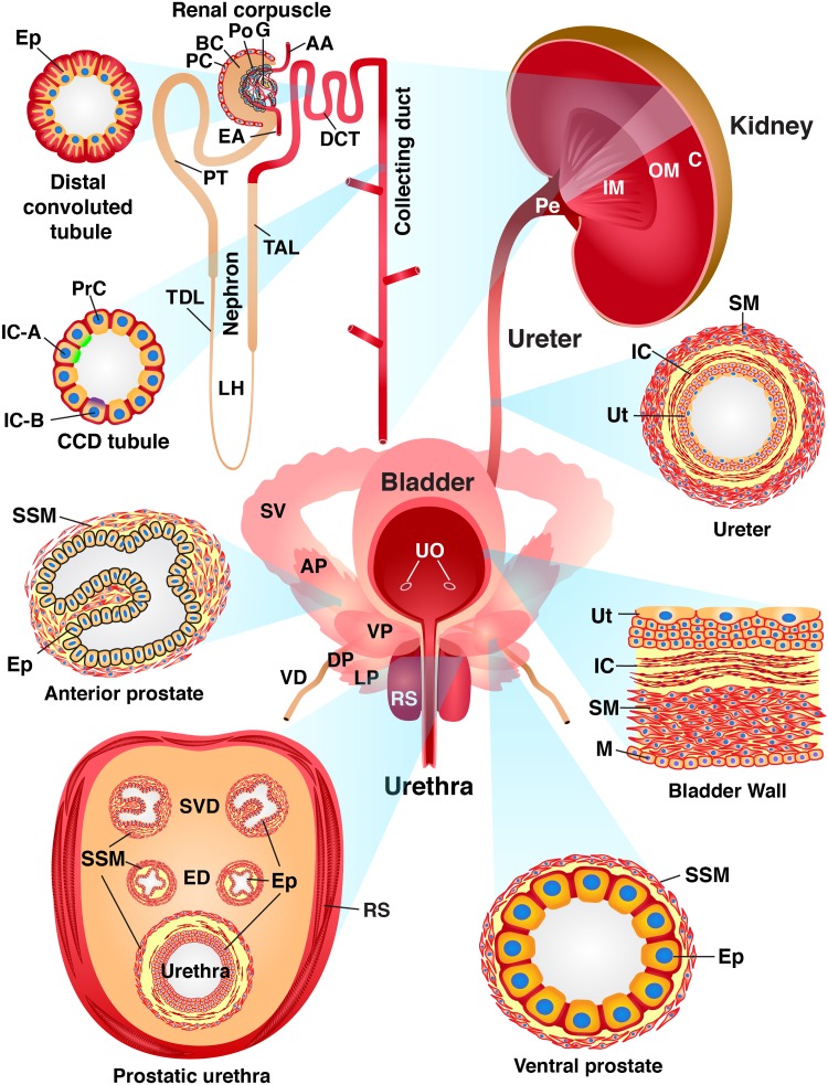
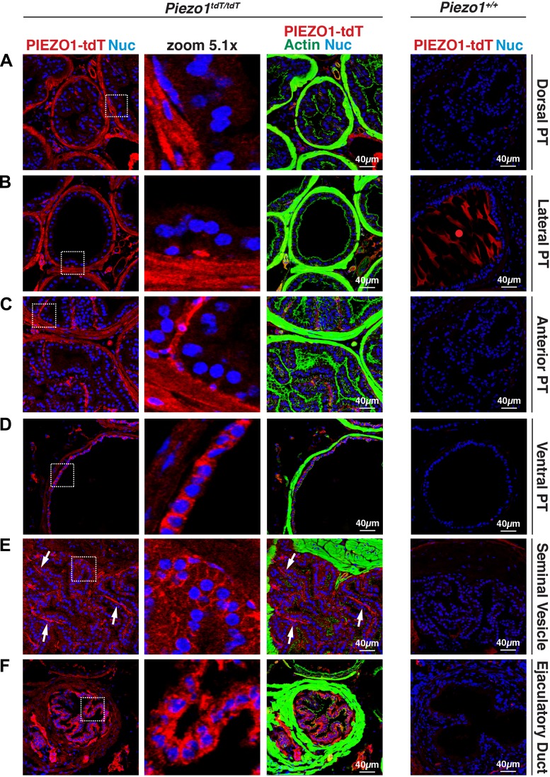
The proper function of the organs that make up the urinary tract (kidneys, ureters, bladder, and urethra) depends on their ability to sense and respond to mechanical forces, including shear stress and wall tension. However, we have limited understanding of the mechanosensors that function in these organs and the tissue sites in which these molecules are expressed. Possible candidates include stretch-activated PIEZO channels (PIEZO1 and PIEZO2), which have been implicated in mechanically regulated body functions including touch sensation, proprioception, lung inflation, and blood pressure regulation. Using reporter mice expressing a COOH-terminal fusion of Piezo1 with the sequence for the tandem-dimer Tomato gene, we found that PIEZO1 is expressed in the kidneys, ureters, bladder, and urethra as well as organs in close proximity, including the prostate, seminal vesicles and ducts, ejaculatory ducts, and the vagina. We further found that PIEZO1 expression is not limited to one cell type; it is observed in the endothelial and parietal cells of the renal corpuscle, the basolateral surfaces of many of the epithelial cells that line the urinary tract, the interstitial cells of the bladder and ureters, and populations of smooth and striated muscle cells. We propose that in the urinary tract, PIEZO1 likely functions as a mechanosensor that triggers responses to wall tension.

Keywords: PIEZO channel; bladder; kidney; mechanotransduction; urinary tract.


Conflict of interest statement

No conflicts of interest, financial or otherwise, are declared by the authors.

Figures

Fig. 1.
Fig. 1.
PIEZO1-tandem-dimer Tomato (tdT) expression in the mouse lung and urinary tract. A: genotype analysis of Piezo1-tdT reporter mice. PCR amplification products of offspring resulting from mating heterozygous Piezo1tdT/+ mice using primers that detect the wild-type allele (227 bp; top) or Piezo1-tdT allele (300 bp; bottom) are shown. B: lung tissue lysate (30 µg) prepared from Piezo1+/+ or Piezo1tdT/tdT mice was resolved by SDS-PAGE and Western blots probed with an antibody against red fluorescent protein (α-RFP; which reacts with the tdT moiety). Detection of β-actin was used as the loading control. C: Western blot analysis of PIEZO1-tdT expression in the different organs of the urinary tract. Tissue lysates from the whole kidney (20 µg), ureters (50 µg), bladder (20 µg), or urethra (20 µg) were prepared from Piezo1+/+ or Piezo1tdT/tdT mice, resolved by PAGE, and detected using Western blot analysis. Values for the relative expression of PIEZO1-tdT in each tissue (normalized to the actin loading control and then expressed as a fraction of the kidney) are indicated below the top set of blots (mean value: n = 2). D: expression of PIEZO1-tdT in the cortex (30 µg) and medulla (10 µg) of Piezo1+/+ and Piezo1tdT/tdT mice kidneys was assessed by Western blots. Values of relative PIEZO1-tdT expression (normalized to the actin loading control and, subsequently, the cortex) are indicated below the top set of blots (mean value: n = 2). E: localization of PIEZO1-tdT in the lung. Tissue obtained from Piezo1+/+ or Piezo1tdT/tdT mice was immunolabeled with antibodies that detect PIEZO1-tdT, platelet endothelial cell adhesion molecule (PECAM)-1, or both markers along with TO-PRO-3. Insets show magnified images of the areas enclosed by the white dashed boxes. Top and upper middle insets show that PIEZO1-tdT was expressed at the apical and basolateral surfaces of the endothelial cells lining blood vessels (surfaces are indicated by arrowheads), whereas the bottom and lower middle insets show that PIEZO1-tdT was also expressed in the alveolar space, including in endothelial cells. L, lumen; MW, molecular weight.
Fig. 2.
Fig. 2.
PIEZO1-tandem-dimer Tomato (tdT) expression and distribution in the mouse kidney. A−E: expression of PIEZO1-tdT in the cortex (A), cortex/outer stripe of the outer medulla (B), inner stripe of the outer medulla (C), inner medulla (D), or renal pelvis (E) of Piezo1tdT/tdT or Piezo1+/+ mice. Tissue was labeled with antibodies that detect PIEZO1-tdT, FITC-phalloidin to label the F-actin cytoskeleton, and TO-PRO-3 to label nuclei. *Renal corpuscles in A. In B, the yellow dashed lines indicate the interface between the cortex (top of image) and outer stripe of the outer medulla (bottom of image). In E, the boxed region of the urothelium lining the renal pelvis is magnified in the inset. Arrows point to the location of the junctional complex, which sits at border between the basolateral and apical plasma membrane domains. The lumen is to the right. The strong F-actin staining (green) is associated with smooth muscle cells that underlie the urothelium.
Fig. 3.
Fig. 3.
Expression of PIEZO1-tandem-dimer Tomato (tdT) in the mouse renal corpuscle but not the proximal tubule. A and B: expression of PIEZO1-tdT in the renal corpuscle of Piezo1tdT/tdT mice immunolabeled with antibodies that detect PIEZO1-tdT and either podocin (NPHS2) or platelet endothelial cell adhesion molecule (PECAM)-1. *Renal corpuscles. Arrows point to the location of parietal cells. Arrowheads point to the location of endothelial cells lining an arteriole. The light gray circle in A is an example of an NPHS2-positive cell whose cytoplasm is PIEZO1-dtT positive. Insets in A show two cells; the left cell expresses PIEZO1-tdT on its surface and is closely associated with the NPHS2-labeled basolateral surface of the adjacent podocyte (see merged image for clarity). The other cell is not PIEZO1-tdT positive and may be associated with a mesangial cell. Insets in B show two PIEZO1-tdT- and PECAM-1-positive endothelial cells. C: expression of PIEZO1-tdT and cubilin (CUBN) in the renal cortex. The CUBN-labeled proximal tubules (indicated by white filled circles) are distinct from the PIEZO1-tdT-labeled tubules (indicated by yellow circles).
Fig. 4.
Fig. 4.
Distribution of PIEZO1-tandem-dimer Tomato (tdT) in the loop of Henle and distal convoluted tubule (DCT) of the mouse kidney. A and B: localization of PIEZO1-tdT, aquaporin-1 (AQP1), and uromodulin (UMOD) in the kidney medulla. AQP1 is a marker of the thin descending limb of the loop of Henle, whereas UMOD is a marker of the thick ascending limb. Inset in A shows the faint PIEZO1-tdT signal associated with the periphery of AQP1-positive tubules, most likely endothelial cells. In B, there was little colocalization between PIEZO1-tdT and UMOD. C and D: localization of PIEZO1-tdT, UMOD, and thiazide-sensitive Na+-Cl cotransporter (SLC12A3) in the kidney cortex. In the cortex, UMOD was associated in part with the early segment of the DCT (i.e., DCT1). In C, the yellow circles indicate PIEZO1-tdT-positive tubules that were negative for UMOD expression. The inset in C shows a section through a UMOD-positive tubule demonstrating the stria formed by PIEZO1-tdT localized to the basolateral surface of the cells lining the DCT. Note the apical position of the nucleus (marked with an arrowhead). In D, colored arrows mark cortical tubules that were positive for UMOD alone (likely thick ascending limb; cyan arrow), SLC12A3 alone (likely segments of late DCT2; white arrow), or positivity for both markers (likely DCT1; yellow arrow). The inset in D shows a region of the tubule lining a segment of the DCT that was SLC12A3 positive (but UMOD negative). Note the PIEZO1-tdT-positive stria at the basolateral surfaces of the cells.
Fig. 5.
Fig. 5.
PIEZO1-tandem-dimer Tomato (tdT) expression in the collecting ducts (CDs) of the mouse kidney. AE: localization of PIEZO1-tdT, aquaporin-2 (AQP2), V-ATPase subunit E1 (ATP6V1E1), and pendrin (SLC26A4) in the kidney. In A, the insets show that PIEZO1-tdT was localized to the basolateral surfaces of AQP2-positive principal cells in the cortical CDs (labeled with yellow circles). B: PIEZO1-tdT colocalized with E-cadherin (CDH1) at the basolateral surfaces of CDs. The inset in C shows that PIEZO1-tdT was associated with the basolateral surfaces of an ATP6V1E1-positive type A intercalated cell, which is distinct from AQP2-positive principal cells. The inset in D shows a cell where PIEZO1-tdT was localized to the basolateral surface of type B interstitial cells expressing apical SLC26A4. The inset in E shows that PIEZO1-tdT was localized to the basolateral surfaces of the cells lining the medullary CD.
Fig. 6.
Fig. 6.
PIEZO1-tandem-dimer Tomato (tdT) expression in the mouse ureter. A−C: distribution of PIEZO1-tdT and platelet-derived growth factor-α receptor (PDGFRA) in the ureters of Piezo1tdT/tdT (A and B) and Piezo1+/+ mice (C). The insets in A show that PIEZO1-tdT was localized to the basolateral surfaces of the outermost umbrella cells that face the lumen. Arrowheads indicate the position of the junctional complex that segregates the apical and basolateral surfaces of these cells. The insets in B show that PDGFRA-positive interstitial cells (one of which is indicated by yellow arrowheads) were PIEZO1-tdT positive. Note that the actin signal in A and C was saturated to reveal the relatively weak signal associated with the urothelium. Ad, adventitia; LP, lamina propria; Nuc, nuclei; SM, smooth muscle; Ut, urothelium.
Fig. 7.
Fig. 7.
PIEZO1-tandem-dimer Tomato (tdT) expression in the mouse bladder. AE: distribution of PIEZO1-tdT in the urothelium (A), lamina propria (B), detrusor (C and D), and mesothelium (E) of Piezo1tdT/tdT and Piezo1+/+ mice. In A, the position of the junctional complex is indicated by arrows. The inset shows a magnified view of a surface umbrella cell labeled with PIEZO1-tdT at its basolateral surfaces. The inset in B shows that PIEZO1-tdT was associated with platelet-derived growth factor-α receptor (PDGFRA)-positive interstitial cells (one of which is indicated by yellow arrowheads). The inset in C shows that PIEZO1-tdT was localized at the sarcolemma of smooth muscle cells. The brighter-stained, stellate-shaped cells, labeled with yellow arrows, are likely intermuscular interstitial cells (see also D). The inset in D shows that PIEZO1-tdT also labeled intermuscular-associated interstitial cells, and the inset in E shows serosa-associated mesothelial cells. Nuc, nuclei.
Fig. 8.
Fig. 8.
PIEZO1-tandem-dimer Tomato (tdT) expression in the mouse urethra. AD: distribution of PIEZO1-tdT in the female urethra (A), female rhabdosphincter (B), female vaginal epithelium (C), or male urethra (D) in Piezo1tdT/tdT and Piezo1+/+ mice. Examples of blood vessels are marked with yellow arrows, and the position of the apicolateral junctional complex is marked with white arrows. The insets in A show the localization of PIEZO1-tdT along the basolateral membranes of surface-localized epithelial cells and the surfaces of the underlying cell layers. The insets in B show that PIEZO1-tdT was expressed by skeletal muscle cells in the rhabdosphincter. The insets in C show the localization of PIEZO1-tdT within the epithelium lining the vagina. The insets in D show the localization of PIEZO1-tdT within the epithelium lining the male urethra. Nuc, nuclei.
Fig. 10.
Fig. 10.
Summary of PIEZO1-tandem-dimer Tomato (tdT) expression in the mouse urinary tract. Cross-sections through the walls of the kidney, ureters, bladder, urethra, and urethra-associated organs are shown to highlight the different cell types that express PIEZO1-tdT (red). The kidney is subdivided into the cortex (C), outer medulla (OM; inner and outer stripe regions not depicted), inner medulla (IM), and pelvis (Pe). The functional units of the kidney include the renal corpuscle [comprised of the following: afferent arteriole (AA), Bowman’s capsule (BC), efferent arteriole (EA), glomerulus (G), parietal cell (PC), and podocyte (Po)], nephron [which includes the distal convoluted tubule (DCT), loop of Henle (LH), proximal tubule (PT), thick ascending limb (TAL), and thin descending limb (TDL)], and collecting ducts (CD). The epithelial cells (Ep) that comprise the DCT characteristically have highly folded basolateral membranes and a subapical nucleus. The cortical CD (CCD) comprises principal cells (PrC), type A intercalated cells [IC-A; which express the V-ATPase E1 subunit (ATP6V1E1) apically], and type B intercalated cells [IC-B; which express pendrin (SLC26A4) apically]. Components of the ureter include the urothelium (Ut), lamina propria-associated interstitial cells (IC), smooth muscle cells (SM), and an adventitia (not depicted). Constituents of the bladder include the urothelium (Ut), interstitial cells in the lamina propria (IC), smooth muscles that form the detrusor (SM), and mesothelial cells (M), which line the serosal surface of the organ. In the male mouse ureter, the prostatic urethra is associated with several structures, including the glands and ducts of the four lobes of the mouse prostate: the anterior prostate (AP), ventral prostate (VP), lateral prostate (LP), and dorsal prostate (DP). Both the AP and VP (as well as the LP and DP, which are not depicted) comprise glands comprised of the epithelium (Ep) surrounded by stromal smooth muscle cells (SSM). In addition to the prostate, the male prostatic urethra is associated with the glands (SV) and ducts (SVD) of the seminal vesicles, the terminal ejaculatory ducts (ED) of the vas deferens (VD), and the surrounding rhabdosphincter (RS), which comprises skeletal muscle. Female mice retain the urethra and rhadbosphincter but lack the male accessory glands. Organs, tissues, and cells not drawn to scale. UO, urethral orifices.
Fig. 9.
Fig. 9.
PIEZO1-tandem-dimer Tomato (tdT) expression in the prostate, seminal vesicles, and ejaculatory ducts of male mice. AF: distribution of PIEZO1-tdT, actin, and nuclei (Nuc) in the dorsal prostate (PT; A), lateral PT (B), anterior PT (C), ventral PT (D), seminal vesicles (E), and ejaculatory ducts (terminal region of ductus deferens; F) of Piezo1tdT/tdT and Piezo1+/+ mice. The actin signal associated with each organ is saturated to show the relatively weak signal associated with the epithelium of these organs. In the dorsal PT, lateral PT, and anterior PT, PIEZO1-tdT was localized primarily to stromal cells surrounding the epithelium. In the ventral PT, PIEZO1-tdT was localized to the stromal cells as well as the basolateral surface of the cuboidal epithelium that lines this gland of the ventral PT. The red circle in B, right, indicates nonspecific staining observed in glands containing large amounts of prostatic secretions. Arrows in E indicate the stromal regions within the papilla that form the seminal vesicles.

References

    1. Albuisson J, Murthy SE, Bandell M, Coste B, Louis-Dit-Picard H, Mathur J, Fénéant-Thibault M, Tertian G, de Jaureguiberry JP, Syfuss PY, Cahalan S, Garçon L, Toutain F, Simon Rohrlich P, Delaunay J, Picard V, Jeunemaitre X, Patapoutian A. Dehydrated hereditary stomatocytosis linked to gain-of-function mutations in mechanically activated PIEZO1 ion channels. Nat Commun 4: 1884, 2013. [Erratum in Nat Commun 4: 2440, 2013.] doi:10.1038/ncomms2899. - DOI - PMC - PubMed
    1. Alper SL. Genetic diseases of PIEZO1 and PIEZO2 dysfunction. Curr Top Membr 79: 97–134, 2017. doi:10.1016/bs.ctm.2017.01.001. - DOI - PubMed
    1. Andolfo I, Alper SL, De Franceschi L, Auriemma C, Russo R, De Falco L, Vallefuoco F, Esposito MR, Vandorpe DH, Shmukler BE, Narayan R, Montanaro D, D’Armiento M, Vetro A, Limongelli I, Zuffardi O, Glader BE, Schrier SL, Brugnara C, Stewart GW, Delaunay J, Iolascon A. Multiple clinical forms of dehydrated hereditary stomatocytosis arise from mutations in PIEZO1. Blood 121: 3925–3935, 2013. doi:10.1182/blood-2013-02-482489. - DOI - PubMed
    1. Apodaca G, Balestreire E, Birder LA. The uroepithelial-associated sensory web. Kidney Int 72: 1057–1064, 2007. doi:10.1038/sj.ki.5002439. - DOI - PubMed
    1. Árnadóttir J, Chalfie M. Eukaryotic mechanosensitive channels. Annu Rev Biophys 39: 111–137, 2010. doi:10.1146/annurev.biophys.37.032807.125836. - DOI - PubMed

Publication types