Review
. 2019 Nov;21(11):e13063.
doi: 10.1111/cmi.13063. Epub 2019 Jun 13.

Molecular interactions between Neisseria meningitidis and its human host

Affiliations
Review

Molecular interactions between Neisseria meningitidis and its human host

Mathieu Coureuil et al. Cell Microbiol. 2019 Nov.

Abstract

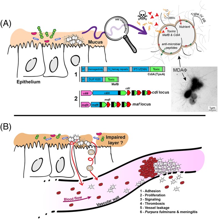
Neisseria meningitidis is a Gram-negative bacterium that asymptomatically colonises the nasopharynx of humans. For an unknown reason, N. meningitidis can cross the nasopharyngeal barrier and invade the bloodstream where it becomes one of the most harmful extracellular bacterial pathogen. This infectious cycle involves the colonisation of two different environments. (a) In the nasopharynx, N. meningitidis grow on the top of mucus-producing epithelial cells surrounded by a complex microbiota. To survive and grow in this challenging environment, the meningococcus expresses specific virulence factors such as polymorphic toxins and MDAΦ. (b) Meningococci have the ability to survive in the extra cellular fluids including blood and cerebrospinal fluid. The interaction of N. meningitidis with human endothelial cells leads to the formation of typical microcolonies that extend overtime and promote vascular injury, disseminated intravascular coagulation, and acute inflammation. In this review, we will focus on the interplay between N. meningitidis and these two different niches at the cellular and molecular level and discuss the use of inhibitors of piliation as a potent therapeutic approach.


Figures

Figure 1
Figure 1
Neisseria meningitidis, a commensal and pathogenic bacterium. A. Nmeningitidis grow in the mucus in nasopharynx where it encounters a poor nutritive medium and a rich microbiota. Meningococci survive by expressing capsule, LOS, the MtrCDE efflux pump, and factors that capture nutrients. Nmeningitidis also express two families of polymorphic toxins: MafB and CdiA. A1 depicts the domain organisation found in CdiA and MafB polymorphic toxins constituted by a conserved N‐terminal domain (blue boxes) apposed to a toxic domain in the variable C‐terminal region (green boxes). Many toxic activities have been reported for toxic domains (Zhang et al., 2012). SP, signal peptide; Hemag activity, hemagglutination activity domain, also called a “TPS domain” (PF05860); fil hemag repeats, filamentous hemagglutinin repeats (PF13332); PT‐VENN, pre‐toxin domain with a VENN motif; DUF1020, domain of unknown function 1020. A2: Simplified genomic organisations of maf and cdi loci. Full‐length toxin genes cdiA and mafB are depicted in blue with their extremity encoding the toxic activity depicted in green. Genes encoding immunity proteins are depicted in red (cdiI and mafI). Open Reading Frame (ORF) encoding alternative C‐terminal toxic domains of CdiA and MafB are depicted in green surrounded with dotted lines. cdiB encodes the dedicated transporter of CdiA toxin, whereas the role of mafA in MafB secretion is unknown. Black boxes indicate regions potentially involved in recombination. Left panel: aggregates harvested from the biomass covering a monolayer of FaDu cells infected by the meningococcal Z5463 strain (Bille et al., 2017) and labelled by the anti‐MDA polyclonal antibody coupled to 8 nm‐diameter gold particles. Representative picture of meningococcal MDAΦ phage‐dependant aggregates. Bar: 1 μm. B. For an unknown reason, meningococci cross the epithelial layer and enter the bloodstream where bacteria adhere to the vascular wall. Adhesive bacteria proliferate and induce an active signalling that leads to better adhesion and opening of the vascular barrier, vessel leakage, and massive thrombosis

References

    1. Ahmed, M. , & Cheung, N. K. (2014). Engineering anti‐GD2 monoclonal antibodies for cancer immunotherapy. FEBS Letters, 588, 288–297. 10.1016/j.febslet.2013.11.030 - DOI - PubMed
    1. Ali, M. Y. (1965). Histology of the human nasopharyngeal mucosa. Journal of Anatomy, 99, 657–672. - PMC - PubMed
    1. Aoki, S. K. , Pamma, R. , Hernday, A. D. , Bickham, J. E. , Braaten, B. A. , & Low, D. A. (2005). Contact‐dependent inhibition of growth in Escherichia coli. Science (New York, N.Y.), 309, 1245–1248. 10.1126/science.1115109 - DOI - PubMed
    1. Arenas, J. , de Maat, V. , Caton, L. , Krekorian, M. , Herrero, J. C. , Ferrara, F. , & Tommassen, J. (2015). Fratricide activity of MafB protein of N. meningitidis strain B16B6. BMC Microbiology, 15, 156 10.1186/s12866-015-0493-6 - DOI - PMC - PubMed
    1. Arenas, J. , Paganelli, F. L. , Rodriguez‐Castano, P. , Cano‐Crespo, S. , van der Ende, A. , van Putten, J. P. , & Tommassen, J. (2016). Expression of the Gene for Autotransporter AutB of Neisseria meningitidis Affects Biofilm Formation and Epithelial Transmigration. Frontiers in Cellular and Infection Microbiology, 6, 162. - PMC - PubMed

MeSH terms