. 2019 Jun 4;8(6):794.
doi: 10.3390/jcm8060794.

Simvastatin Attenuates Cardiac Fibrosis via Regulation of Cardiomyocyte-Derived Exosome Secretion

Affiliations

Simvastatin Attenuates Cardiac Fibrosis via Regulation of Cardiomyocyte-Derived Exosome Secretion

Hsuan-Fu Kuo et al. J Clin Med. .

Abstract

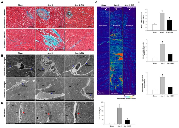
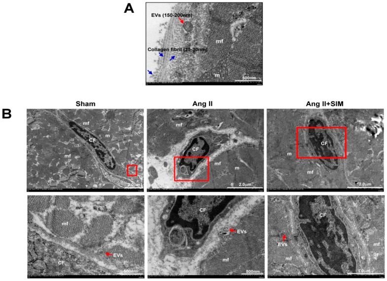
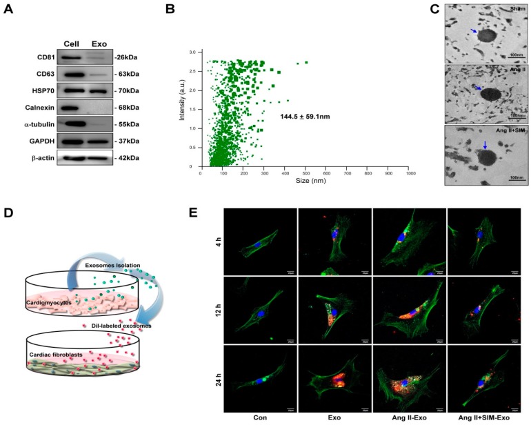
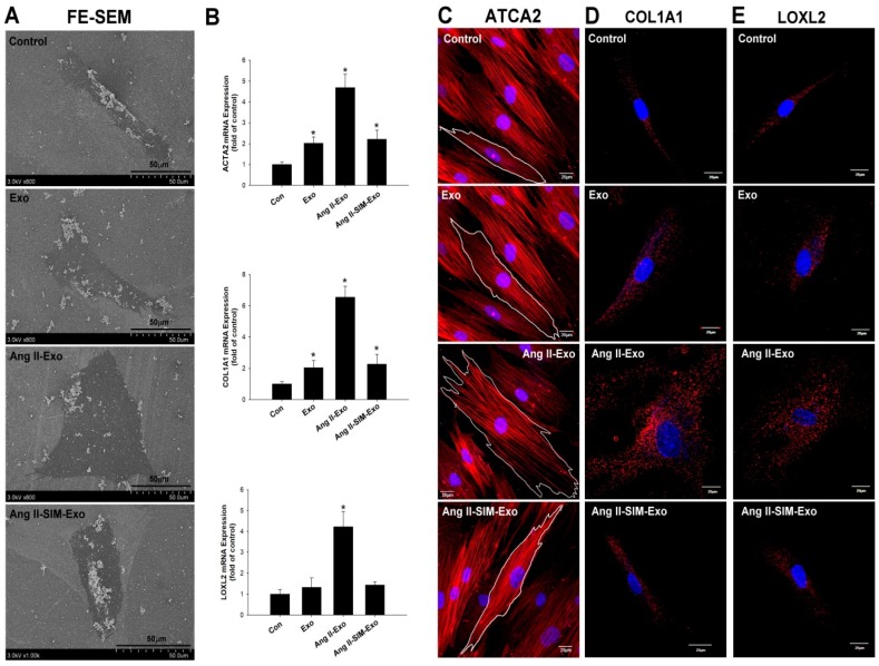
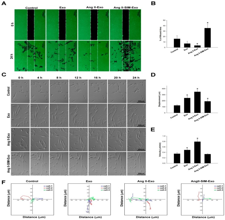
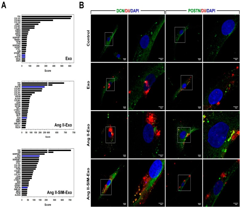
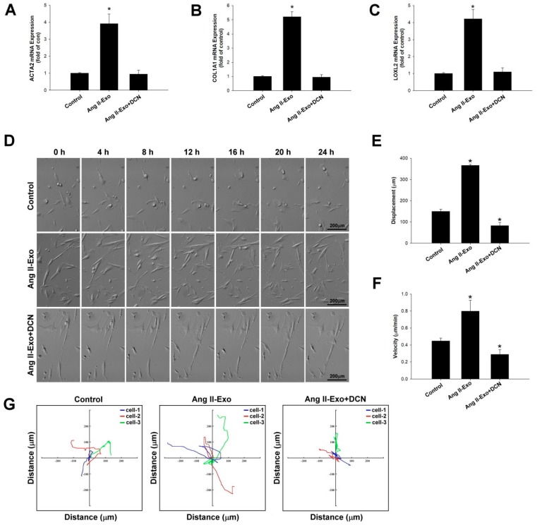
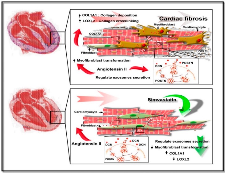
Exosome-mediated communication within the cardiac microenvironment is associated with cardiac fibrosis. Simvastatin (SIM), a potent statin, protects against cardiac fibrosis, but its mechanism of action is unclear. We investigated the inhibitory effects and underlying mechanism of simvastatin in cardiac fibrosis, by regulating exosome-mediated communication. Male Sprague-Dawley rats were treated with angiotensin (Ang) II alone, or with SIM for 28 d. Cardiac fibrosis, expressions of fibrosis-associated proteins and mRNAs, and collagen fiber arrangement and deposition were examined. Protein expressions in exosomes isolated from Ang II-treated cardiomyocytes (CMs) were evaluated using nano-ultra-performance liquid chromatographic system, combined with tandem mass spectrometry. Transformation of fibroblasts to myofibroblasts was evaluated using scanning electron and confocal microscopy, and migration assays. Our results showed that SIM attenuated in vivo expression of collagen and collagen-associated protein, as well as collagen deposition, and cardiac fibrosis. The statin also upregulated decorin and downregulated periostin in CM-derived exosomes. Furthermore, it suppressed Ang II-induced transformation of fibroblast to myofibroblast, as well as fibroblast migration. Exosome-mediated cell-cell communication within the cardiac tissue critically regulated cardiac fibrosis. Specifically, SIM regulated the release of CM exosomes, and attenuated Ang II-induced cardiac fibrosis, highlighting its potential as a novel therapy for cardiac fibrosis.

Keywords: cardiac fibrosis; decorin; exosomes; periostin; statins.


Conflict of interest statement

The authors declare no conflicts of interest.

Figures

Figure 1
Figure 1
Simvastatin suppresses in vivo angiotensin (Ang) II-mediated collagen deposition, fibrosis, and collagen-associated protein expression. Male Sprague-Dawley rats were treated with (Ang II, 1 mg/kg/day) or Ang II + simvastatin (SIM, oral, 10 mg/kg) for 28 days. Perivascular and interstitial fibrosis ultrastructures (blue color) were determined using Masson’s trichrome staining. (A) The collagen fibers are shown in the representative images (blue) (n = 6). Perivascular and interstitial collagen fiber expression (blue arrows) (B) and quantification by transmission electron microscopy (TEM) analysis are shown (Supplemental Figure S1A,B). The cardiac fibroblast (CF) ultrastructures (red arrows) were determined using TEM analysis (C) (n = 6) (mf, myofibril; m, mitochondrial; CF, cardiac fibroblast). Cardiac collagen deposition and arrangement from endocardium to epicardium were identified and quantified using second-harmonic generation (SHG) microscopy (D) (n = 3). * p < 0.05 vs. the control from three independent experiments. The fibrotic associated genes: ACTA2, COL1A1, and LOXL2 expression were analyzed using real-time PCR and normalized by β–actin (E). For all comparisons, * p < 0.05 vs. sham.
Figure 2
Figure 2
Ultrastructure between CM and CF of the heart in vivo. The SD rats were divided into sham group, Ang II group and Ang II + SIM group (each group n = 6). TEM images of rat left ventricles demonstrated the morphology, size of longitudinal and cross-sectional collagen fibers, and extracellular vesicles (EVs) (A). The distribution of EVs between CM and CF was explored by TEM analysis (B) (mf, myofibril; m, mitochondrial; CF, cardiac fibroblast).
Figure 3
Figure 3
HCM-derived exosomes are transported into human cardiac fibroblasts (HCF) in vitro. Exosomal-specific marker expression from HCM-isolated exosomes was measured using Western blot analysis (A). HCM-isolated exosome size was demonstrated by NanoSight NS300 (B) and TEM analysis (C). To identify uptake of HCM-derived exosomes by HCF, exosomes were isolated from HCM-conditioned medium, after treatment with Ang II, or Ang II + SIM for 24 h. HCM exosomes were labeled with the DiI dye (5 μg/mL, red florescence) for 1 h, and incubated with HCF cells for various times (4, 12, and 24 h) (D). HCF cells were non-treated (control), or treated with Eox, Ang II-Exo and Ang II-SIM-Exo for various times (4, 12, and 24 h), then stained for intracellular cytoskeleton F-actin (green florescence; nucleus, blue florescence), and analyzed by confocal microscopy (E). Scale bars: 20 μm. Each image is representative of three independent experiments.
Figure 4
Figure 4
Simvastatin attenuates myofibroblast phenotype transformation and collagen-associated protein expression in vitro. Exosomes were isolated from HCM culture medium pretreated with Ang II or Ang II + SIM for 24 h. Exosomes were then incubated with HCF for 24 h. HCF ultrastructure images were examined using scanning electron microscopy (SEM) (A). mRNA expressions of myofibroblast markers ATCA2, COL1A1, and LOXL2 were examined by real-time PCR and normalized by β–actin (B). Data represent mean ± SD of three independent experiments (* p < 0.05). Confocal microscopy was used to confirm ATCA2, COL1A1, and LOXL2 expressions (red florescence: ATCA2, COL1A1, LOXL2; blue florescence, the nuclei) (C–E). A representative image from three independent experiments.
Figure 5
Figure 5
Simvastatin inhibits HCF migration after induction with HCM-derived exosomes in vitro. Exosomes were isolated from the culture medium of HCM cells pretreated with Ang II or Ang II + SIM for 24 h, and then HCF cells were incubated with human HCF cells for 24 h. HCF cell migration mediated by HCM-derived exosomes was analyzed (A), and quantified (B) by wound healing assay. Scale bar, 500 µm. * p < 0.05 vs. control from three independent experiments. To reveal simvastatin inhibition of HCF cell mobility, time-lapse confocal microscopy was used. Images were acquired (C), cell movements were measured, and displacement (μm) (D), velocity (μm/min) (E), and motility tracks (x and y axis; distance μm) (F) were quantified. * p < 0.05 vs. control from three independent experiments.
Figure 6
Figure 6
Simvastatin regulates HCM-derived exosome protein content in vitro. HCM-derived exosome proteins were isolated after Ang II or Ang II+SIM treatment and identified using nano UPLC-MS/MS (A). To confirm exosomal decorin (DCN) and periostin (POSTN) protein expressions, HCM-derived exosomes were labeled with DiI dye (5 μg/mL, red florescence) for 1 h, and then incubated with HCF cells for 24 h. Immunofluorescence staining was performed (DCN and POSTN, green fluorescence; exosomes-labeled with DiI dye, red fluorescence; nucleus, blue fluorescence) in HCF cells (B). A representative image from three independent experiments is shown.
Figure 7
Figure 7
DCN inhibits collagen-associated gene expression, fibroblast transformation, and cell motility in vitro. The effects of DCN on ATCA2, COL1A1, and LOXL2 expressions were examined using real-time PCR and normalized by β–actin (AC). * p < 0.05 vs. control from three independent experiments. DCN effect on HCF cell migration was examined using time-lapse confocal microscopy. Images were acquired (D), and cell movements measured as displacement (μm) (E), velocity (μm/min) (F), and motility tracks (x and y axis; distance μm) (G), were plotted. * p < 0.05 vs. control from three independent experiments.
Figure 8
Figure 8
Suggested mechanism underlying Simvastatin-induced suppression of Ang II-mediated fibroblast. SIM might suppress fibroblast to myofibroblast transformation, collagen production/deposition, and increased fibroblast motility by inducing cardiomyocyte secretion of exosomal proteins.

Similar articles

Cited by

References

    1. Schnee J.M., Hsueh W.A. Angiotensin II, adhesion, and cardiac fibrosis. Cardiovasc. Res. 2000;46:264–268. doi: 10.1016/S0008-6363(00)00044-4. - DOI - PubMed
    1. Wang Q., Yu Y., Zhang P., Chen Y., Li C., Chen J., Wang Y., Li Y. The crucial role of activin A/ALK4 pathway in the pathogenesis of Ang-II-induced atrial fibrosis and vulnerability to atrial fibrillation. Basic Res. Cardiol. 2017;112:47. doi: 10.1007/s00395-017-0634-1. - DOI - PubMed
    1. Fan Z., Guan J. Antifibrotic therapies to control cardiac fibrosis. Biomater. Res. 2016;20:13. doi: 10.1186/s40824-016-0060-8. - DOI - PMC - PubMed
    1. Zhang P., Su J., Mende U. Cross talk between cardiac myocytes and fibroblasts: From multiscale investigative approaches to mechanisms and functional consequences. Am. J. Physiol. Heart Circ. Physiol. 2012;303:H1385–H1396. doi: 10.1152/ajpheart.01167.2011. - DOI - PMC - PubMed
    1. Kofron C.M., Mende U. In vitro models of the cardiac microenvironment to study myocyte and non-myocyte crosstalk: Bioinspired approaches beyond the polystyrene dish. J. Physiol. 2017;595:3891–3905. doi: 10.1113/JP273100. - DOI - PMC - PubMed

LinkOut - more resources