Review
. 2019 Jun 5;83(3):e00007-19.
doi: 10.1128/MMBR.00007-19. Print 2019 Aug 21.

Gut Microbiota and Colonization Resistance against Bacterial Enteric Infection

Affiliations
Review

Gut Microbiota and Colonization Resistance against Bacterial Enteric Infection

Q R Ducarmon et al. Microbiol Mol Biol Rev. .

Abstract

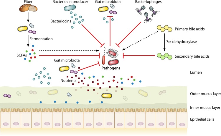
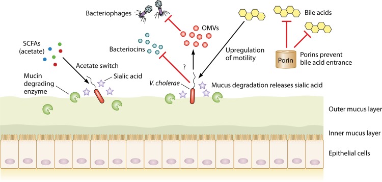
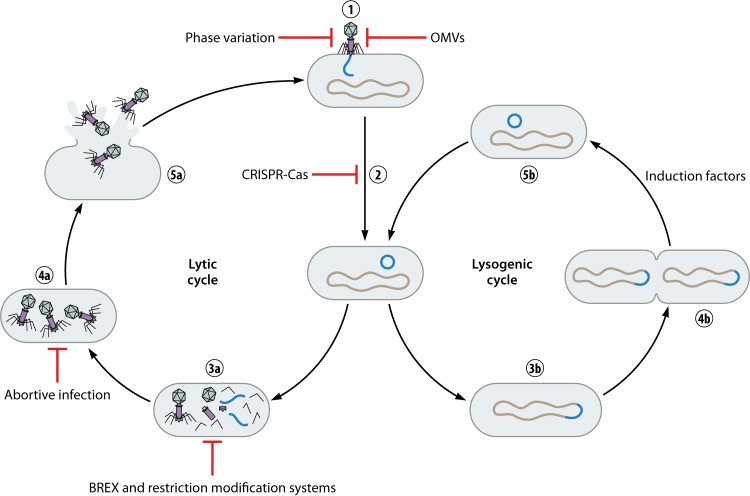
The gut microbiome is critical in providing resistance against colonization by exogenous microorganisms. The mechanisms via which the gut microbiota provide colonization resistance (CR) have not been fully elucidated, but they include secretion of antimicrobial products, nutrient competition, support of gut barrier integrity, and bacteriophage deployment. However, bacterial enteric infections are an important cause of disease globally, indicating that microbiota-mediated CR can be disturbed and become ineffective. Changes in microbiota composition, and potential subsequent disruption of CR, can be caused by various drugs, such as antibiotics, proton pump inhibitors, antidiabetics, and antipsychotics, thereby providing opportunities for exogenous pathogens to colonize the gut and ultimately cause infection. In addition, the most prevalent bacterial enteropathogens, including Clostridioides difficile, Salmonella enterica serovar Typhimurium, enterohemorrhagic Escherichia coli, Shigella flexneri, Campylobacter jejuni, Vibrio cholerae, Yersinia enterocolitica, and Listeria monocytogenes, can employ a wide array of mechanisms to overcome colonization resistance. This review aims to summarize current knowledge on how the gut microbiota can mediate colonization resistance against bacterial enteric infection and on how bacterial enteropathogens can overcome this resistance.

Keywords: bacterial enteric infection; bacteriocins; bacteriophages; bile acids; colonization resistance; enteric pathogens; gut microbiota; microbiome; mucus layer; nutrient competition; short-chain fatty acids.


Figures

FIG 1
FIG 1
Outline of gut microbiota-mediated colonization resistance mechanisms. Fiber obtained from the diet is fermented by the gut microbiota into SCFAs. Bacteriocin producers produce bacteriocins capable of targeting a specific pathogen. Primary bile acids can be converted by a very select group of gut microbiota into secondary bile acids, which generally have properties antagonistic to pathogens. Nutrient competition of native microbiota can limit access to nutrients for a pathogen. Specific organisms can use SCFAs, bacteriocins, and primary bile acids to increase their virulence, as discussed in the text.
FIG 2
FIG 2
V. cholerae uses a wide array of mechanisms to overcome CR. First, it employs its acetate switch to use acetate to upregulate its own virulence. Nothing about potential bacteriocin resistance is presently known, and this subject remains to be studied. To protect itself from bacteriophages, V. cholerae produces outer membrane vesicles (OMVs) that act as a decoy binding site for the attacking phages (see Bacterial Defense Mechanisms against Bacteriophages). Regulation of outer membrane porins allows them to prevent entry of bile acids when they are encountered. By employing specific mucin-degrading enzymes, V. cholerae releases sialic acid and subsequently metabolizes it.
FIG 3
FIG 3
Lytic and lysogenic bacteriophage infection cycles with bacterial defense mechanisms. The first two steps of infection are identical for the lytic and lysogenic cycles, namely, phage binding followed by DNA insertion and DNA circularization (1 and 2). The lysogenic cycle then branches off by integrating its DNA into the bacterial chromosome and becoming a prophage, thereby ensuring its replication (3b). Only upon encountering induction factors will the prophage leave the bacterial chromosome, after which it can enter the lytic cycle (4b and 5b). In the lytic cycle, phage DNA and protein are replicated and subsequently assembled into full phages (3a and 4a). The phages then lyse the bacterial cell, are released, and can infect other bacteria (5a). Bacteria possess multiple mechanisms to prevent killing by bacteriophages, starting with blocking attachment. This can be achieved through phase variation or production of OMVs. After phage DNA entry, CRISPR-Cas can recognize this foreign DNA and degrade it. Phage DNA and protein replication can be prevented by BREX and restriction-modification systems, while full phage assembly can be prevented by abortive infection.

References

    1. Sender R, Fuchs S, Milo R. 2016. Revised estimates for the number of human and bacteria cells in the body. PLoS Biol 14:e1002533. doi:10.1371/journal.pbio.1002533. - DOI - PMC - PubMed
    1. Sommer F, Backhed F. 2013. The gut microbiota—masters of host development and physiology. Nat Rev Microbiol 11:227–238. doi:10.1038/nrmicro2974. - DOI - PubMed
    1. Round JL, Palm NW. 2018. Causal effects of the microbiota on immune-mediated diseases. Sci Immunol 3:eaao1603. doi:10.1126/sciimmunol.aao1603. - DOI - PubMed
    1. Opazo MC, Ortega-Rocha EM, Coronado-Arrazola I, Bonifaz LC, Boudin H, Neunlist M, Bueno SM, Kalergis AM, Riedel CA. 2018. Intestinal microbiota influences non-intestinal related autoimmune diseases. Front Microbiol 9:432. doi:10.3389/fmicb.2018.00432. - DOI - PMC - PubMed
    1. Li Q, Gao Z, Wang H, Wu H, Liu Y, Yang Y, Han L, Wang X, Zhao L, Tong X. 2018. Intestinal immunomodulatory cells (T lymphocytes): a bridge between gut microbiota and diabetes. Mediators Inflamm 2018:9830939. doi:10.1155/2018/9830939. - DOI - PMC - PubMed

Publication types

Substances