. 2019 Jul 30;93(16):e00250-19.
doi: 10.1128/JVI.00250-19. Print 2019 Aug 15.

Hepatitis B Virus X Protein Function Requires Zinc Binding

Affiliations

Hepatitis B Virus X Protein Function Requires Zinc Binding

Dhivya Ramakrishnan et al. J Virol. .

Abstract

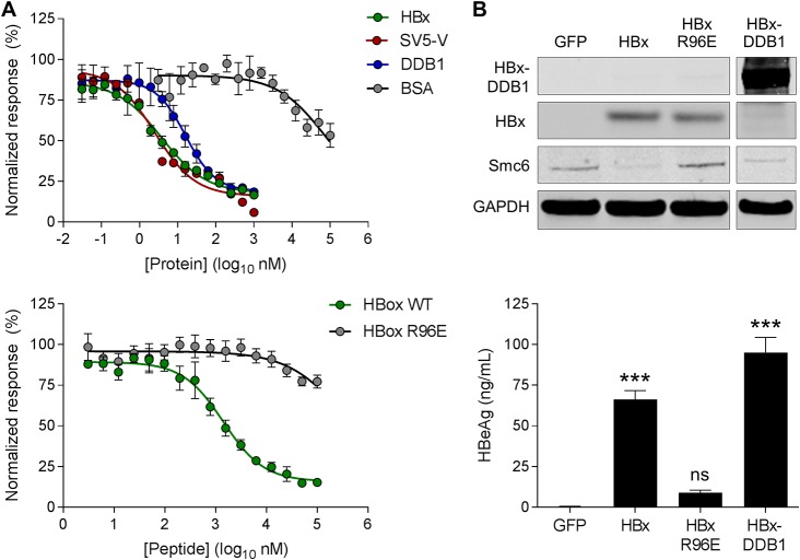
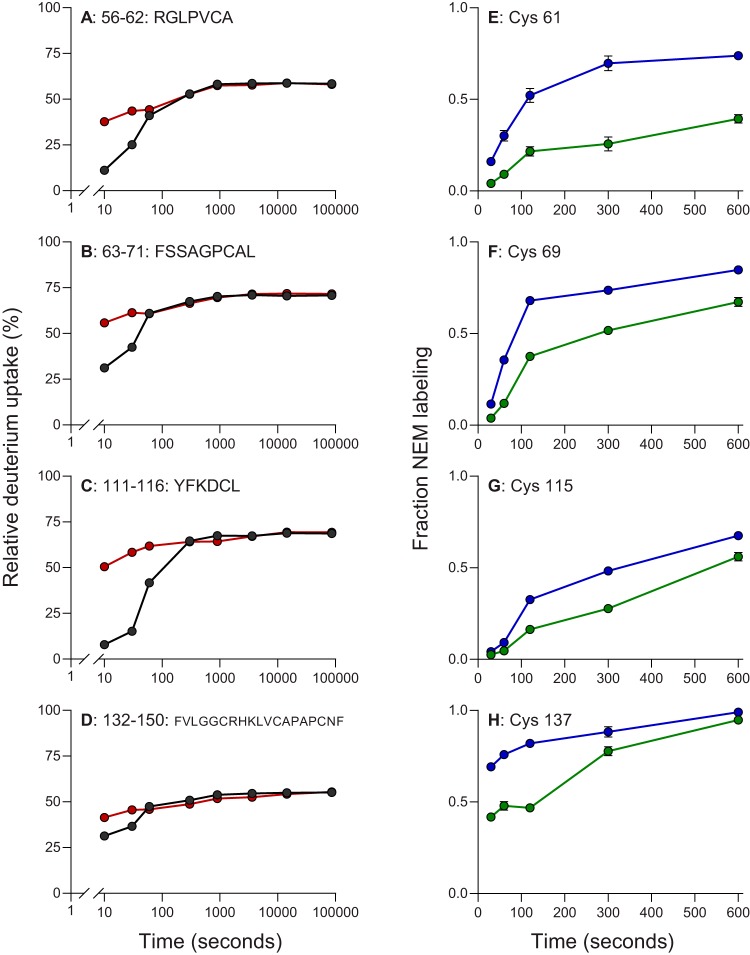
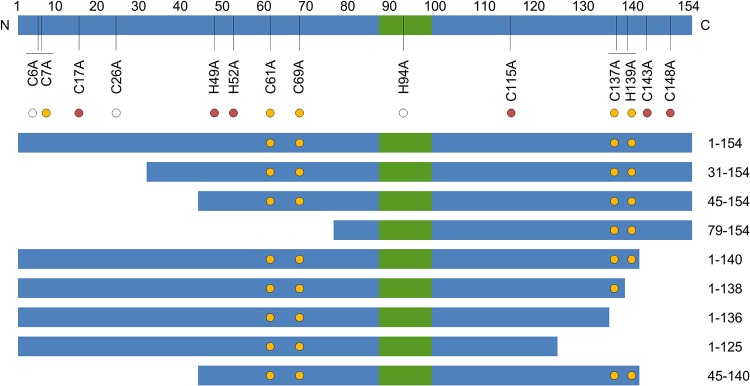
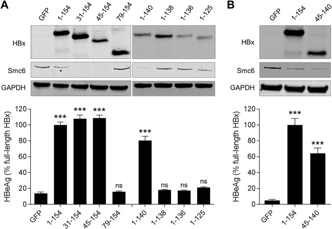
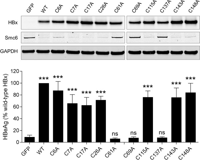
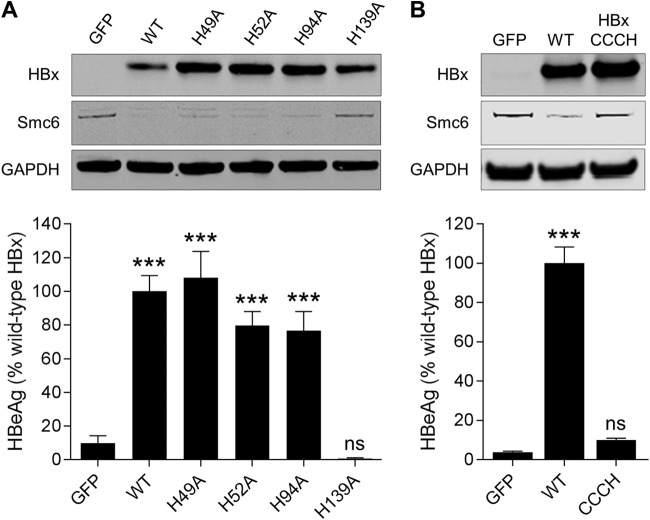
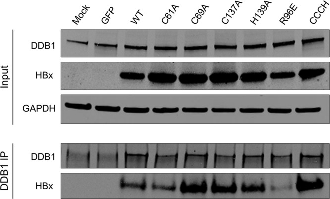
The host structural maintenance of chromosomes 5/6 complex (Smc5/6) suppresses hepatitis B virus (HBV) transcription. HBV counters this restriction by expressing the X protein (HBx), which redirects the cellular DNA damage-binding protein 1 (DDB1)-containing E3 ubiquitin ligase to target Smc5/6 for degradation. However, the details of how HBx modulates the interaction between DDB1 and Smc5/6 remain to be determined. In this study, we performed biophysical analyses of recombinant HBx and functional analysis of HBx mutants in HBV-infected primary human hepatocytes (PHH) to identify key regions and residues that are required for HBx function. We determined that recombinant HBx is soluble and exhibits stoichiometric zinc binding when expressed in the presence of DDB1. Mass spectrometry-based hydrogen-deuterium exchange and cysteine-specific chemical footprinting of the HBx:DDB1 complex identified several HBx cysteine residues (located between amino acids 61 and 137) that are likely involved in zinc binding. These cysteine residues did not form disulfide bonds in HBx expressed in human cells. In line with the biophysical data, functional analysis demonstrated that HBx amino acids 45 to 140 are required for Smc6 degradation and HBV transcription in PHH. Furthermore, site-directed mutagenesis determined that C61, C69, C137, and H139 are necessary for HBx function, although they are likely not essential for DDB1 binding. This CCCH motif is highly conserved in HBV as well as in the X proteins from various mammalian hepadnaviruses. Collectively, our data indicate that the essential HBx cysteine and histidine residues form a zinc-binding motif that is required for HBx function.IMPORTANCE The structural maintenance of chromosomes 5/6 complex (Smc5/6) is a host restriction factor that suppresses HBV transcription. HBV counters this restriction by expressing HBV X protein (HBx), which redirects a host ubiquitin ligase to target Smc5/6 for degradation. Despite this recent advance in understanding HBx function, the key regions and residues of HBx required for Smc5/6 degradation have not been determined. In the present study, we performed biochemical, biophysical, and cell-based analyses of HBx. By doing so, we mapped the minimal functional region of HBx and identified a highly conserved CCCH motif in HBx that is likely responsible for coordinating zinc and is essential for HBx function. We also developed a method to produce soluble recombinant HBx protein that likely adopts a physiologically relevant conformation. Collectively, this study provides new insights into the HBx structure-function relationship and suggests a new approach for structural studies of this enigmatic viral regulatory protein.

Keywords: DDB1; HBx; Smc5/6 complex; hepatitis B virus.


Figures

FIG 1
FIG 1
Characterization of recombinant HBx proteins. (A, top) TR-FRET analysis of the coexpressed His-HBx:GST-DDB1 complex in the presence of recombinant untagged HBx, SV5-V, DDB1, and BSA. (Bottom) TR-FRET analysis of the coexpressed His-HBx:GST-DDB1 complex in the presence of the wild-type (WT) HBx H-box peptide and a mutant HBx R96E H-box peptide. (B, top) Western blot analysis of PHH lysates collected 72 h after transduction with lentiviruses expressing GFP, Myc-HBx, Myc-HBx R96E, or the Myc-HBx-DDB1 fusion. HBx and the HBx-DDB1 fusion were detected using an anti-Myc antibody. Data are representative of results from 3 independent experiments. (Bottom) PHH were transduced with the same lentiviruses and 1 day later were infected with HBVΔX. Extracellular HBeAg levels at day 13 postinfection are expressed as a percentage of the value for the HBx control; the bar height indicates the mean of data from 3 independent experiments, and the error bars represent the SEM. Statistical significance relative to the GFP control was calculated by one-way ANOVA with Dunnett’s multiple-comparison correction. ***, P < 0.001; ns, not significant (P > 0.05).
FIG 2
FIG 2
Identification of HBx cysteine residues protected upon zinc binding using HDX and kinetic NEM footprinting. (A to D) Comparison of deuterium uptake kinetic plots for select HBx peptides in the coexpressed HBx:DDB1 complex in the presence (black) and absence (red) of zinc. These peptides are representative of larger regions in the protein that show changes in HDX upon binding (as described in the text). (E to H) Kinetics of NEM labeling of select cysteine residues of HBx protein in the coexpressed HBx:DDB1 complex in the presence (green) and absence (blue) of zinc. The circles indicate the means, and the error bars represent the SEM. Statistical significance was calculated by a two-tailed t test. Only the four cysteines with significant differences (P < 0.01) are shown.
FIG 3
FIG 3
HBx mutants evaluated in PHH. Shown is a simplified schematic representation of the HBx protein; the green box denotes the H-box. HBx residue numbers are shown directly above the schematic, and HBx point mutations are displayed directly below. The circle color represents the degree of sequence conservation for each residue. The white circle denotes <90% sequence conservation in HBx. The red circle signifies >90% sequence conservation in HBx but not conservation in woodchuck, woolly monkey, and bat hepadnaviruses. The yellow circle denotes >90% sequence conservation in HBx and conservation in the woodchuck, woolly monkey, and bat hepadnaviruses. Truncation mutants are shown below the schematic, with the N and C termini of each mutant displayed on the right. The yellow circles in each HBx truncation mutant represent the positions (if present) of C61, C69, C137, and H139.
FIG 4
FIG 4
Identification of the regions required for HBx function in HBV-infected PHH. (A, top) PHH were transduced with lentiviruses expressing GFP, full-length HBx (HBx1–154), or HBx with either an N-terminal or C-terminal truncation. Cell lysates were analyzed by Western blotting at 72 h posttransduction. HBx was detected using an anti-Myc antibody. (Bottom) PHH were transduced with the same lentiviruses and 1 day later were infected with HBVΔX. Extracellular HBeAg was measured at day 13 postinfection. (B) PHH were transduced with lentiviruses expressing GFP, full-length HBx (HBx1–154), or HBx45–140. Cell lysates were analyzed by Western blotting at 72 h posttransduction. HBx was detected using an anti-Myc antibody. (Bottom) PHH were transduced with the same lentiviruses and 1 day later were infected with HBVΔX. Extracellular HBeAg was measured at day 13 postinfection. Data are expressed as a percentage of the value for the full-length HBx control; the bar height indicates the mean of data from 3 independent experiments, and the error bars represent the SEM. Statistical significance relative to the GFP control was calculated by one-way ANOVA with Dunnett’s multiple-comparison correction. ***, P < 0.001; ns, not significant (P > 0.05).
FIG 5
FIG 5
Identification of cysteine and histidine residues required for HBx function in HBV-infected PHH. (Top) PHH were transduced with lentiviruses expressing GFP, wild-type HBx (WT), or HBx mutants in which a single cysteine residue was mutated to alanine. Cell lysates were analyzed by Western blotting at 72 h posttransduction. HBx was detected using an anti-Myc antibody. (Bottom) PHH were transduced with the same lentiviruses and 1 day later were infected with HBVΔX. Extracellular HBeAg was measured at day 13 postinfection. Data are expressed as a percentage of the value for the wild-type HBx control; the bar height indicates the mean of data from 3 independent experiments, and the error bars represent the SEM. Statistical significance relative to the GFP control was calculated by one-way ANOVA with Dunnett’s multiple-comparison correction. ***, P < 0.001; ns, not significant (P > 0.05).
FIG 6
FIG 6
Identification of histidine residues required for HBx function in HBV-infected PHH. (A, top) PHH were transduced with lentiviruses expressing GFP, wild-type HBx (WT), or HBx mutants in which a single histidine residue was mutated to alanine. Cell lysates were analyzed by Western blotting at 72 h posttransduction. HBx was detected using an anti-Myc antibody. (Bottom) PHH were transduced with the same lentiviruses and 1 day later were infected with HBVΔX. Extracellular HBeAg was measured at day 13 postinfection. (B, top) PHH were transduced with lentiviruses expressing GFP, wild-type HBx, or HBx in which cysteine 61 (C61), C69, C137, and histidine 139 (H139) were all mutated to alanine (HBxCCCH). Cell lysates were analyzed by Western blotting at 72 h posttransduction. HBx was detected using an anti-Myc antibody. (Bottom) PHH were transduced with the same lentiviruses and 1 day later were infected with HBVΔX. Extracellular HBeAg was measured at day 13 postinfection. Data are expressed as a percentage of the value for the wild-type HBx control; the bar height indicates the mean of data from 2 independent experiments, and the error bars represent the SEM. Statistical significance relative to the GFP control was calculated by one-way ANOVA with Dunnett’s multiple-comparison correction. ***, P < 0.001; ns, not significant (P > 0.05).
FIG 7
FIG 7
HBx C61, C69, C137, and H139 are not required for DDB1 binding. PHH were transduced with lentiviruses expressing the indicated proteins. (Top) Cell lysates were analyzed by Western blotting at 72 h posttransduction. (Bottom) DDB1 immunoprecipitation (IP) of the cell lysates was analyzed by Western blotting. HBx was detected using an anti-Myc antibody. Data are representative of results from 3 independent experiments.

References

    1. Schweitzer A, Horn J, Mikolajczyk RT, Krause G, Ott JJ. 2015. Estimations of worldwide prevalence of chronic hepatitis B virus infection: a systematic review of data published between 1965 and 2013. Lancet 386:1546–1555. doi:10.1016/S0140-6736(15)61412-X. - DOI - PubMed
    1. Lozano R, Naghavi M, Foreman K, Lim S, Shibuya K, Aboyans V, Abraham J, Adair T, Aggarwal R, Ahn SY, Alvarado M, Anderson HR, Anderson LM, Andrews KG, Atkinson C, Baddour LM, Barker-Collo S, Bartels DH, Bell ML, Benjamin EJ, Bennett D, Bhalla K, Bikbov B, Bin Abdulhak A, Birbeck G, Blyth F, Bolliger I, Boufous S, Bucello C, Burch M, Burney P, Carapetis J, Chen H, Chou D, Chugh SS, Coffeng LE, Colan SD, Colquhoun S, Colson KE, Condon J, Connor MD, Cooper LT, Corriere M, Cortinovis M, de Vaccaro KC, Couser W, Cowie BC, Criqui MH, Cross M, Dabhadkar KC, et al. . 2012. Global and regional mortality from 235 causes of death for 20 age groups in 1990 and 2010: a systematic analysis for the Global Burden of Disease Study 2010. Lancet 380:2095–2128. doi:10.1016/S0140-6736(12)61728-0. - DOI - PMC - PubMed
    1. Kornyeyev D, Voitenleitner C, Livingston CM, Xing W, Hung M, Kwon HJ, Fletcher SP, Beran RK. 2019. Spatiotemporal analysis of hepatitis B virus X protein in primary human hepatocytes. J Virol 93:e00248-19. doi:10.1128/JVI.00248-19. - DOI - PMC - PubMed
    1. Lucifora J, Arzberger S, Durantel D, Belloni L, Strubin M, Levrero M, Zoulim F, Hantz O, Protzer U. 2011. Hepatitis B virus X protein is essential to initiate and maintain virus replication after infection. J Hepatol 55:996–1003. doi:10.1016/j.jhep.2011.02.015. - DOI - PubMed
    1. Riviere L, Gerossier L, Ducroux A, Dion S, Deng Q, Michel ML, Buendia MA, Hantz O, Neuveut C. 2015. HBx relieves chromatin-mediated transcriptional repression of hepatitis B viral cccDNA involving SETDB1 histone methyltransferase. J Hepatol 63:1093–1102. doi:10.1016/j.jhep.2015.06.023. - DOI - PubMed

Publication types