. 2019 Jun 18;116(25):12343-12352.
doi: 10.1073/pnas.1901080116. Epub 2019 Jun 5.

On the occurrence of cytochrome P450 in viruses

Affiliations

On the occurrence of cytochrome P450 in viruses

David C Lamb et al. Proc Natl Acad Sci U S A. .

Abstract

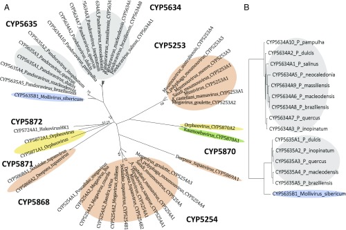
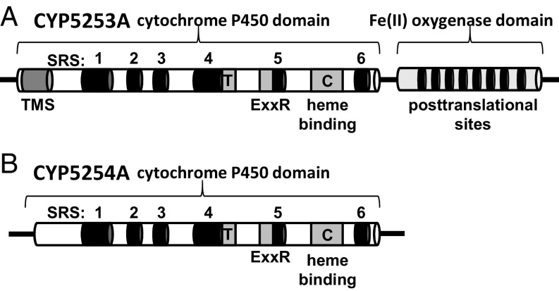
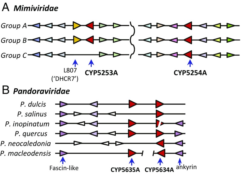
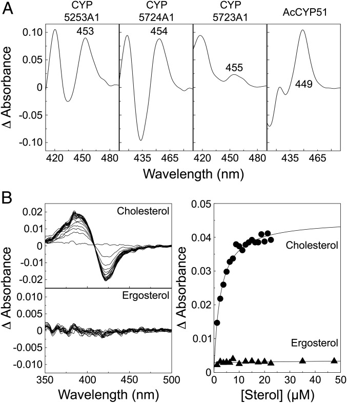
Genes encoding cytochrome P450 (CYP; P450) enzymes occur widely in the Archaea, Bacteria, and Eukarya, where they play important roles in metabolism of endogenous regulatory molecules and exogenous chemicals. We now report that genes for multiple and unique P450s occur commonly in giant viruses in the Mimiviridae, Pandoraviridae, and other families in the proposed order Megavirales. P450 genes were also identified in a herpesvirus (Ranid herpesvirus 3) and a phage (Mycobacterium phage Adler). The Adler phage P450 was classified as CYP102L1, and the crystal structure of the open form was solved at 2.5 Å. Genes encoding known redox partners for P450s (cytochrome P450 reductase, ferredoxin and ferredoxin reductase, and flavodoxin and flavodoxin reductase) were not found in any viral genome so far described, implying that host redox partners may drive viral P450 activities. Giant virus P450 proteins share no more than 25% identity with the P450 gene products we identified in Acanthamoeba castellanii, an amoeba host for many giant viruses. Thus, the origin of the unique P450 genes in giant viruses remains unknown. If giant virus P450 genes were acquired from a host, we suggest it could have been from an as yet unknown and possibly ancient host. These studies expand the horizon in the evolution and diversity of the enormously important P450 superfamily. Determining the origin and function of P450s in giant viruses may help to discern the origin of the giant viruses themselves.

Keywords: cytochrome P450; domains of life; evolution; redox partner; virus.


Conflict of interest statement

The authors declare no conflict of interest.

Figures

Fig. 1.
Fig. 1.
Megavirales phylogeny constructed from the RNAPol2 gene. This ML phylogenetic tree shows the relationship of key members of the Megavirales viruses. The clades that contain encoded cytochrome P450 genes are highlighted in colors that correspond to the cytochrome P450 phylogeny in Fig. 2. Bootstrap support values are derived from 250 replicates, using the HIVB (HIV between-patient) model of amino acid substitution. Only values below 90% are shown.
Fig. 2.
Fig. 2.
Molecular phylogeny of virus cytochrome P450 genes. (A) Unrooted ML phylogeny of predicted amino acid sequences for viral P450s shows the close relationships between Pandoraviridae CYP5634 and CYP5635, and among Mimiviridae CYP5253s and CYP5254s. Note the CYP5254s in tupanviruses, and the CYP5870s from the Kaumoebavirus and Orpheovirus. (B) Detailed phylogeny of selected Pandoraviridae CYP5634s and CYP5635s and Mollivirus CYP5635. Color blocks are the same as in Fig. 1.
Fig. 3.
Fig. 3.
Domain regions in Mimivirus CYP5353A1 (A) and CYP5254A1 (B). C, absolute conserved cysteine residue present in the heme binding domain; ExxR, conserved glutamate-residue-residue arginine motif found in the K helix; SRS, substrate recognition site; T, conserved threonine in the P450 I helix; TMS, transmembrane segment. PTMs include one N-glycosylation site, one protein kinase C phosphorylation site, four caesin kinase II phosphorylation sites, and three myristoylation sites.
Fig. 4.
Fig. 4.
Microsynteny around giant virus cytochrome P450 genes. (A) Mimiviridae: groups A, B, and C display high degrees of shared gene order extending 10 genes around both CYP5253A (Left) and CYP5254A (Right). Genes that are highly similar to each other are colored the same across the various genomes. Completely shared synteny is evident across all three clades for CYP5254A. Clade C viruses are missing both CYP5253A (L808 ortholog) and gene R807 [a putative 7-dehydrocholesterol reductase (DHCR7)]. (B) Pandoraviridae: Note shared synteny around P450s in pandoraviruses in clades A and B. The figure also shows that CYP5634 and CYP5635 are adjacent, and illustrates that CYP5634A1 may be a pseudogene in P. inopinatum. Genes in white are not similar, while purple-colored genes are orthologous. Other identifiable genes are indicated (fascin-like domain and ankyrin).
Fig. 5.
Fig. 5.
Characterization of recombinant cytochromes P450. (A) Dithionite-reduced, CO difference spectra of the purified viral cytochromes P450 and the control A. castellanii CYP51 (AcCYP51) were obtained. The concentrations of P450 used were as follows: CYP5253A1 (2.5 μM), CYP5724A1 (2.5 μM), CYP5723A1 (1.25 μM), and AcCYP51 (1.25 μM), respectively. (B) P450-sterol binding assays: Stock solutions of 2.5 mM cholesterol (●) and ergosterol (▲) were titrated against 4 μM CYP5253A1 in 50 mM Tris⋅HCl (pH 8) and 20% (vol/vol) glycerol, with the difference spectrum determined after each addition of sterol, which were used to construct ligand saturation curves.
Fig. 6.
Fig. 6.
Structural features of CYP102L1. (A) Dithionite-reduced CO difference spectra of the purified Adler phage CYP102L1 (P450 concentration = 1.25 μM) showing a Soret spectral maximum at 448 nm. (B) Open crystal structure of CYP102L1 (Protein Data Bank ID code 6N6Q) exhibits the typical P450-fold consisting of α-helical and β-sheet domains as seen in all other known P450 structures. Heme is shown as a red stick model. (C and D) Comparison of the crystal structures of Adler phage CYP102L1 (green) and B. megaterium CYP102A1 (P450 BM3; blue). Although sequence identity remains low (34%), a number of residues that have been implicated in CYP102A1 function are conserved between both proteins, including A82 (92), F87 (97), F261 (275), A264 (278), T268 (282), A328 (245), A330 (347), F393 (412), and I401 (420), with CYP102L1 residues in parentheses. The CYP102A1 residues are involved in substrate binding affinity and turnover (A82 and I401), substrate selectivity (F87, T268, A328, and A330), and electron transfer (F261, A264, T268, and F393). Additional residues and references documenting residue involvement are cited in SI Appendix.

References

    1. Klingenberg M., Pigments of rat liver microsomes. Arch. Biochem. Biophys. 75, 376–386 (1958). - PubMed
    1. Omura T., Sato R., A new cytochrome in liver microsomes. J. Biol. Chem. 237, 1375–1376 (1962). - PubMed
    1. Estabrook R. W., Cooper D. Y., Rosenthal O., The light reversible carbon monoxide inhibition of the steroid C21-hydroxylase system of the adrenal cortex. Biochem. Z. 338, 741–755 (1963). - PubMed
    1. Nelson D. R., Goldstone J. V., Stegeman J. J., The cytochrome P450 genesis locus: The origin and evolution of animal cytochrome P450s. Philos. Trans. R. Soc. Lond. B Biol. Sci. 368, 20120474 (2013). - PMC - PubMed
    1. Woese C. R., Fox G. E., Phylogenetic structure of the prokaryotic domain: The primary kingdoms. Proc. Natl. Acad. Sci. U.S.A. 74, 5088–5090 (1977). - PMC - PubMed

Publication types

MeSH terms

Substances

Associated data