

IRRITANT AND ALLERGIC CONTACT DERMATITIS - SKIN LESION CHARACTERISTICS
- PMID: 31168208
- PMCID: PMC6544100
- DOI: 10.20471/acc.2018.57.04.13
IRRITANT AND ALLERGIC CONTACT DERMATITIS - SKIN LESION CHARACTERISTICS
Abstract
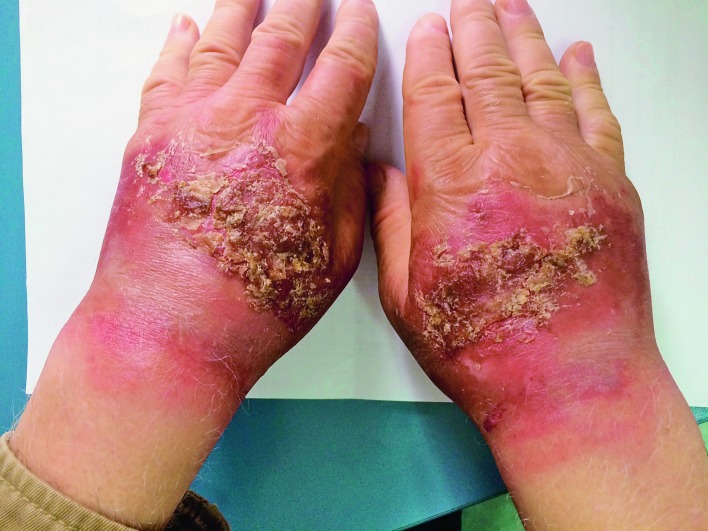
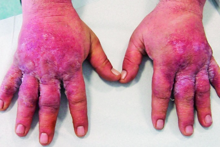
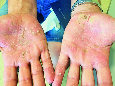
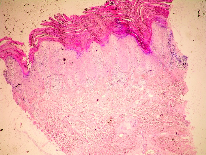
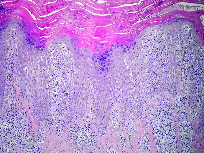
- Contact skin lesions may be the consequences of contact with various irritants or allergens, or due to other factors (e.g., UV radiation, microbials), intrinsic factors (e.g., in autoimmune responses), or even their combination. There are many substances related to irritant contact dermatitis (CD), causing irritant or toxic effects, e.g., chemical and physical agents, plants, phototoxic agents, airborne irritants, etc. Impaired barrier function (e.g., aberrancies in epidermal pH buffering capabilities) also participates by promoting bacterial biofilms and creating an environment favoring sensitization. Development of allergic CD skin lesions includes complex immune pathways and inflammatory mediators, influenced by both genetic (predominantly filaggrin mutations) and environmental triggers. In the pathogenesis of allergic CD, antimicrobial peptides play a prominent role; they are produced by various skin cells (e.g., keratinocytes, sebocytes) and move to inflamed lesions during an inflammation process. Also, in allergic CD skin lesions, the skin shows different types of immune responses to individual allergens, although clinical manifestations do not depend on the causative allergen type, e.g., nickel stimulates immune activation primarily of the Th1/Th17 and Th22 components. Also important are alarmins, proteases, immunoproteomes, lipids, natural moisturizing factors, tight junctions, smoking, etc. We expect that future perspectives may reveal new pathogenetic factors and scientific data important for the workup and treatment of patients with CD.
Keywords: Dermatitis, allergic contact; Dermatitis, irritant; Etiopathogenesis; Factors; Histology; Immunohistochemistry; Skin inflammation.
Figures
References
-
- Nair PA, Atwater AR. Dermatitis, Contact. [Updated 2017 Oct 4]. In: StatPearls [Internet]. Treasure Island (FL): StatPearls Publishing; 2017 Jun-. Available from: https://www.ncbi.nlm.nih.gov/books/NBK459230/
-
- Przybilla B, Rueff F. Contact dermatitis. In: Burgdorf WHC, Plewig G, Wolf HH, Landthaler M. Braun-Falco’s Dermatology. Berlin: Springer-Verlag; 2009. p. 377-401.
-
- Lugović L, Šitum M, Ožanić-Bulić S, Sjerobabski-Masnec I. Phototoxic and photoallergic skin reactions. Coll Antropol. 2007;31 Suppl 1:63–7. - PubMed
Publication types
MeSH terms
Substances
LinkOut - more resources
Full Text Sources