. 2019 Mar 27;4(2):344-350.
doi: 10.1002/epi4.12317. eCollection 2019 Jun.

Therapeutic effect of Anakinra in the relapsing chronic phase of febrile infection-related epilepsy syndrome

Affiliations

Therapeutic effect of Anakinra in the relapsing chronic phase of febrile infection-related epilepsy syndrome

Robertino Dilena et al. Epilepsia Open. .

Abstract

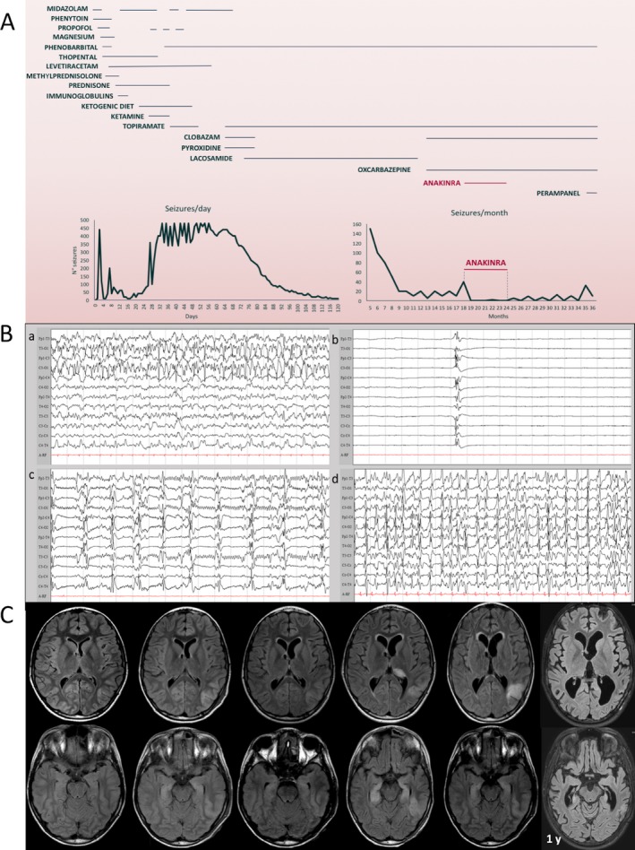
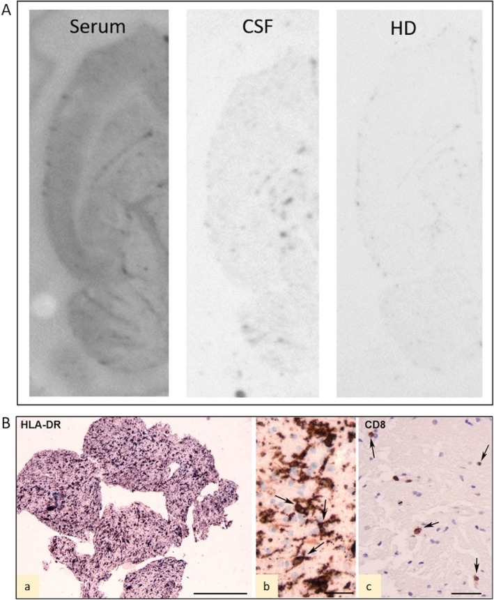
Febrile infection-related epilepsy syndrome (FIRES) is a severe epileptic encephalopathy with presumed inflammatory origin and lacking effective treatments. Anakinra is the human recombinant interleukin 1 receptor antagonist clinically used in autoinflammatory or autoimmune conditions. We report a case of FIRES for which the spatial and temporal match between electroencephalography (EEG) and magnetic resonance imaging (MRI) focal alterations provides support for the detrimental synergic interplay between seizures and inflammation that may evolve to permanent focal lesions and progressive brain atrophy in weeks to months. Brain biopsy showed aspects of chronic neuroinflammation with scarce parenchymal lymphocytes. We report the novel evidence that anakinra reduces the relapse of highly recurrent refractory seizures at 1.5 years after FIRES onset. Our evidence, together with previously reported therapeutic effects of anakinra administered since the first days of disease onset, support the hypothesis that interleukin 1β and inflammation-related factors play a crucial role in seizure recurrence in both the acute and chronic stages of the disease.

Keywords: IL‐1β; epileptic encephalopathy; febrile infection–related epilepsy syndrome; neuroinflammation.


Conflict of interest statement

None of the authors has any conflict of interest to disclose. All co‐authors have been substantially involved in the study and/or the preparation of the manuscript; no undisclosed groups or persons have had a primary role in the study and/or in manuscript preparation. All co‐authors have seen and approved the submitted version of the paper and accept responsibility for its content. We confirm that we have read the Journal's position on issues involved in ethical publication and affirm that this report is consistent with those guidelines.

Figures

Figure 1
Figure 1
Evolution of the disease and treatment effect. A, Trend of seizures and treatments during the 3 years after disease onset. Thiopental sedation in the acute phase induces burst‐suppression with status epilepticus rebound at thiopental withdrawal. During the chronic phase, seizure reduction is attained by anakinra. B, Representative electroencephalography (EEG) tracings acquired during the first month from disease onset. a: EEG tracing of the second day during a left temporal‐occipital epileptic activity; b: burst‐suppression EEG pattern induced by high‐dose thiopental used to control the aggressive status epilepticus; c: relapse of epileptic abnormalities upon reduction of thiopental sedation; d: status epilepticus after interruption of continuous pharmacologic burst‐suppression (26 days from disease onset). C, Brain magnetic resonance imaging (MRI) evolution during the first year from disease onset. The first brain MRI performed at 2 days (d) from the onset of status epilepticus was unremarkable. The second and third MRI studies at 9 and 21 days showed a stable cortical hyperintensity in the left temporal‐occipital region, with a slight hyperintensity of both hippocampi. The fourth MRI (41 day) was performed after more than 10 days from burst‐suppression interruption (indicated by dotted vertical line in C) with the patient experiencing rebound of aggressive ongoing multifocal and generalized seizures. A more evident left temporal‐occipital hyperintensity involved not only the cortical region, but also the subcortical white matter. A diffuse hyperintensity of the bilateral hippocampal region and of the pulvinar region of the left thalamus became evident. In the fifth MRI study (78 days) after gradual reduction of epileptic activity, the hippocampal alteration was slightly reduced and the pulvinar alteration was no longer present, whereas the temporal‐occipital lesion was still present and a progressive increase in ventricular size became evident. At 1‐year follow‐up, the MRI showed a global brain atrophy with an increased enlargement of the posterior temporal horn. The left temporal‐occipital hyperintensity as well as the hippocampal hyperintensity were no longer visible, but these structures were deeply atrophic
Figure 2
Figure 2
Immunoblots and histopathology. A, Immunohistoblots showing the labeling of horizontal transversal hemisections of rat brain with patient's serum and cerebrospinal fluid (CSF). No labeling was observed in sections incubated with a pool of healthy donor (HD) sera. The principal brain structure identified by patient's serum is neocortex (see Ref. 8). B, Histopathology of biopsy of left posterior temporal lobe. The biopsy consists of small fragments of predominant white matter (Wm) with presence of activated HLA‐DR‐positive cells (a); high magnification photographs of HLA‐DR‐positive reactive microglia within the Wm (arrows in b); few scattered CD8‐positive T lymphocytes (arrows in c). Scale bars: A, 1 mm; B, 25 μm; C, 40 μm. Anti‐human leukocyte antigen (HLA)‐DP, DQ, DR (HLA‐DR; clone CR3/43) anti‐CD8 (mouse monoclonal, clone C8/144B)

References

    1. Hirsch LJ, Gaspard N, van Baalen A, Nabbout R, Demeret S, Loddenkemper T, et al. Proposed consensus definitions for new‐onset refractory status epilepticus (NORSE), febrile infection‐related epilepsy syndrome (FIRES), and related conditions. Epilepsia 2018;59:739–744. - PubMed
    1. Vezzani A, Maroso M, Balosso S, Sanchez MA, Bartfai T. IL‐1 receptor/Toll‐like receptor signaling in infection, inflammation, stress and neurodegeneration couples hyperexcitability and seizures. Brain Behav Immun 2011;25:1281–1289. - PubMed
    1. Kenney‐Jung DL, Vezzani A, Kahoud RJ, LaFrance‐Corey RG, Ho ML, Muskardin TW, et al. Febrile infection‐related epilepsy syndrome treated with anakinra. Ann Neurol 2016;80:939–945. - PMC - PubMed
    1. Jyonouchi H, Geng L. Intractable Epilepsy (IE) and Responses to Anakinra, a Human Recombinant IL‐1 Receptor Antagonist (IL‐1Ra): case Reports. J Clin Cell Immunol 2016;7:456–460.
    1. DeSena AD, Do T, Schulert GS. Systemic autoinflammation with intractable epilepsy managed with interleukin‐1 blockade. J Neuroinflammation 2018;15:38. - PMC - PubMed