

[Expression of autophagy-related protein Beclin-1 and microtubule-associated protein 2 light chain 3 in periodontal ligament cells in orthodontic tooth pressure areas]
- PMID: 31168983
- PMCID: PMC7030146
- DOI: 10.7518/hxkq.2019.02.008
[Expression of autophagy-related protein Beclin-1 and microtubule-associated protein 2 light chain 3 in periodontal ligament cells in orthodontic tooth pressure areas]
Abstract
Objective: To investigate the expression of autophagy-related protein Beclin-1 and microtubule-associated protein 2 light chain 3 (LC3Ⅱ) in periodontal ligament cells in orthodontic tooth pressure areas.
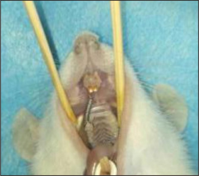
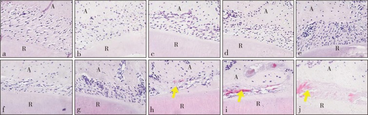
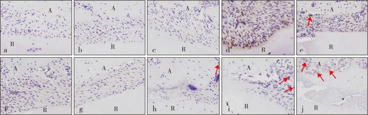
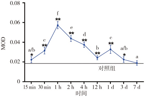
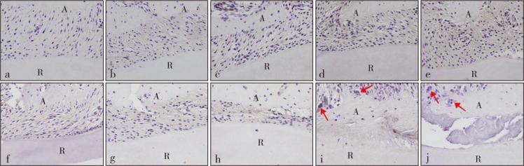
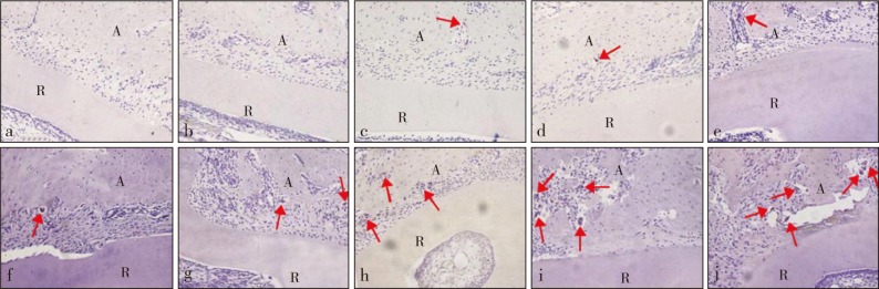
Methods: Sixty male SD rats were randomly divided into a blank control group and nine experimental groups. In the experimental groups, 0.392 N orthodontic force was used to move the first right upper molars for 15 min, 30 min, 1 h, 2 h, 4 h, 12 h, 1 d, 3 d, or 7 d. The blank control group did not receive any treatment. The rats were euthanized. Changes in the morphology of the periodontal membrane in the pressure areas were observed through hematoxylin and eosin (HE) staining. The expression levels of Beclin-1 and LC3Ⅱ were detected by immunohistochemical staining, and tartrate-resistant acid phosphatase (TRAP) staining was performed for the counting of osteoclasts.
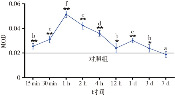
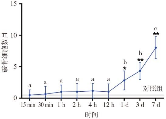
Results: The HE stains showed that the hyalinization of the periodontal ligament appeared in the pressure areas after 1 day of exertion and was gradually aggravated. The immunohistochemical stains showed that the expression levels of Beclin-1 and LC3Ⅱ in the experimental groups gradually increased, peaked after 1 h, and then gradually decreased. The expression levels peaked again after 1 d, then decreased to baseline levels at 7 d of exertion. Beclin-1 and LC3Ⅱ were expressed in the osteoclasts. The TRAP stains indicated that the number of osteoclasts started to increase after 1 day.
Conclusions: Autophagy may participate in the process of periodontal ligament reconstruction in orthodontic tooth pressure areas by mediating the hyalinization of periodontal ligament and affecting the biological effects of osteoclasts.
目的 探究正畸牙压力区牙周膜细胞自噬相关蛋白Beclin-1与微管相关蛋白2轻链3(LC3Ⅱ)的表达。方法 将60只雄性SD大鼠随机分为空白对照组和9个实验组,实验组正畸加力0.392 N近中移动右上第一磨牙,加力时间分别为15 min、30 min、1 h、2 h、4 h、12 h、1 d、3 d、7 d,空白对照组不做任何处理。处死大鼠后,行苏木精-伊红(HE)染色观察压力区牙周膜形态学变化、免疫组织化学染色检测Beclin-1与LC3Ⅱ的表达、抗酒石酸酸性磷酸酶(TRAP)染色计数破骨细胞。结果 HE染色显示,加力1 d后压力区牙周膜透明样变出现,并随加力时间延长逐渐加重。免疫组织化学染色显示,实验组Beclin-1和LC3Ⅱ表达均上调,1 h达峰值,随后逐渐降低,1 d时再次增强达一小峰值,后又回降,7 d时降低至基线水平。破骨细胞中也可见Beclin-1和LC3Ⅱ的表达。TRAP染色提示,加力1 d后破骨细胞数量开始增加。结论 自噬或许通过介导牙周膜透明样变发生和影响破骨细胞生物学作用参与正畸牙压力区牙周膜改建的过程。.
Keywords: autophagy; orthodontic tooth movement; periodontal ligament; pressure.
Figures
References
-
- Dutra EH, Nanda R, Yadav S. Bone response of loaded periodontal ligament[J] Curr Osteoporos Rep. 2016;14(6):280–283. - PubMed
-
- Ma KG, Shao ZW, Yang SH, et al. Autophagy is activated in compression-induced cell degeneration and is mediated by reactive oxygen species in nucleus pulposus cells exposed to compression[J] Osteoarthritis Cartilage. 2013;21(12):2030–2038. - PubMed
-
- Baskaran R, Poornima P, Priya LB, et al. Neferine prevents autophagy induced by hypoxia through activation of Akt/mTOR pathway and Nrf2 in muscle cells[J] Biomed Pharmacother. 2016;83:1407–1413. - PubMed
-
- Huang ZJ, Fang WL, Liu WH, et al. Aspirin induces Beclin-1-dependent autophagy of human hepatocellular carcinoma cell[J] Eur J Pharmacol. 2018;823:58–64. - PubMed
MeSH terms
Substances
LinkOut - more resources
Full Text Sources