. 2019 May 21:10:1111.
doi: 10.3389/fimmu.2019.01111. eCollection 2019.

Molecular Characterization of Monocyte Subsets Reveals Specific and Distinctive Molecular Signatures Associated With Cardiovascular Disease in Rheumatoid Arthritis

Collaborators, Affiliations

Molecular Characterization of Monocyte Subsets Reveals Specific and Distinctive Molecular Signatures Associated With Cardiovascular Disease in Rheumatoid Arthritis

Patricia Ruiz-Limon et al. Front Immunol. .

Abstract

Objectives: This study, developed within the Innovative Medicines Initiative Joint Undertaking project PRECISESADS framework, aimed at functionally characterize the monocyte subsets in RA patients, and analyze their involvement in the increased CV risk associated with RA. Methods: The frequencies of monocyte subpopulations in the peripheral blood of 140 RA patients and 145 healthy donors (HDs) included in the PRECISESADS study were determined by flow cytometry. A second cohort of 50 RA patients and 30 HDs was included, of which CD14+ and CD16+ monocyte subpopulations were isolated using immuno-magnetic selection. Their transcriptomic profiles (mRNA and microRNA), proinflammatory patterns and activated pathways were evaluated and related to clinical features and CV risk. Mechanistic in vitro analyses were further performed. Results: CD14++CD16+ intermediate monocytes were extended in both cohorts of RA patients. Their increased frequency was associated with the positivity for autoantibodies, disease duration, inflammation, endothelial dysfunction and the presence of atheroma plaques, as well as with the CV risk score. CD14+ and CD16+ monocyte subsets showed distinctive and specific mRNA and microRNA profiles, along with specific intracellular signaling activation, indicating different functionalities. Moreover, that specific molecular profiles were interrelated and associated to atherosclerosis development and increased CV risk in RA patients. In vitro, RA serum promoted differentiation of CD14+CD16- to CD14++CD16+ monocytes. Co-culture with RA-isolated monocyte subsets induced differential activation of endothelial cells. Conclusions: Our overall data suggest that the generation of inflammatory monocytes is associated to the autoimmune/inflammatory response that mediates RA. These monocyte subsets, -which display specific and distinctive molecular signatures- might promote endothelial dysfunction and in turn, the progression of atherosclerosis through a finely regulated process driving CVD development in RA.

Keywords: cardiovascular disease; gene profile; microRNAs; monocyte subsets; rheumatoid arthritis.


Figures
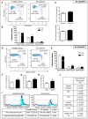
Figure 1
Figure 1
Relationship of the increased intermediate monocyte subpopulation in RA patients with their immunologic and inflammatory profile, and association to endothelial dysfunction, CIMT and CV risk score. (A–C) PRECISESADS Cohort. (A) Representative dot plots of monocyte subtypes from RA patients and HDs. (B) Percentage of different monocyte subtypes by flow cytometry in whole blood of 140 RA patients and 145 HDs. (C) Association between increased frequency of intermediate monocytes and autoimmunity. (D–J) Second cohort: 50 RA patients and 33 HDs. (D) Representative dot plots of monocyte subtypes from RA patients and HDs. (E) Percentage of different monocyte subtypes by flow cytometry. (F–H) Association between increased frequency of intermediate monocytes with autoimmunity and pathological CIMT. (I) Impaired microvascular endothelial dysfunction in RA patients measured by Laser-Doppler. (J) Correlations between the percentage of intermediate monocytes and clinical parameters of the disease, endothelial dysfunction and SCORE CVD. Paired t-test was performed *indicates significant differences vs. HDs (p < 0.05).
Figure 2
Figure 2
Different protein expression of pro-thrombotic and inflammatory factors in RA monocyte subtypes. (A–D) Protein expression of Tissue factor (TF), IκB kinase (IKK), Tumor necrosis factor (TNF) and interleukin 6 (IL-6) in the three subpopulations of monocytes in RA and HDs measured by flow cytometry. Data are presented as mean ± SD, n = 50 RA patients and 33 HDs. Paired t-test was performed aindicates significant differences vs. HDs, bindicates significant differences vs. CD14++CD16 RA monocytes (p < 0.05).
Figure 3
Figure 3
Specific and differential expression profile of genes related to atherosclerosis in CD16+ and CD14+ monocytes from RA patients compared to HDs. (A) Atherosclerosis RT2 profiler PCR array performed in CD14+ and CD16+ monocytes isolated from 12 RA patients and 12 HDs. Heat-map of the differentially expressed genes in CD16+ RA or CD14+ RA monocytes vs. CD16+ HD and CD14+ HD monocytes, respectively. (B) Validation of the PCR array in CD14+ and CD16+ monocytes through RT-PCR in samples of 50 RA patients and 30 HDs, separately. Kruskal-Wallis test, followed by a Dunn's multiple comparison test was performed. aIndicates significant differences vs. CD14+ HDs, bindicates significant differences vs. CD14+ RA, cindicates significant differences vs. CD16+ HDs (p < 0.05).
Figure 4
Figure 4
CD14+ and CD16+ monocytes of RA patients display specific and distinctive microRNA expression profiles. (A) Thirty-one microRNAs were found significantly altered in CD14+ monocytes isolated from RA patients compared with CD14+ HD monocytes using a fold change cut-off of >2. (B) Functional classification of the altered microRNAs in CD14+ RA monocytes using Ingenuity Pathway Analysis (IPA, QIAGEN Redwood City, https://analysis.ingenuity.com). The analysis included only the functions and pathways with average IPA score >2 [indicated as –log (p-value)].To minimize false positives among significantly enriched functions, a false discovery rate (FDR) < 0,05 (–log P-value = 1,33) was used to determine the probability that each biological function assigned to that data set was due to chance alone'. Threshold bar indicates cut-off point of significance (p > 0.05), using Fisher's exact test, microRNAs. (C) One hundred and seventy-three microRNAs were found significantly altered in CD16+ monocytes isolated from RA patients compared with CD16+ HD monocytes using a fold change cut-off of >2. (D) Functional classification of the altered microRNAs in CD16+ RA monocytes using Ingenuity Pathway Analysis. The analysis included only the functions and pathways with average IPA score >2 [indicated as –log (p-value)]. Threshold bar indicates cut-off point of significance (p >0.05), using Fisher's exact test, microRNAs.
Figure 5
Figure 5
Validation of the microRNA array and integrated analysis between the microRNAs and genes altered in CD14+ and CD16+ RA monocytes. (A) mRNA relative expression of microRNAs in CD14+ and CD16+ monocytes from RA patients and HDs. Data are presented as mean ± SD, n = 50 RA patients and 33 HDs. Paired t-test was performed (*) indicates significant differences vs. HDs (p < 0.05). (B) Integrated analysis of altered microRNAs (microRNA array) and mRNA gene expression (PCR array) in CD14+ RA monocytes. (C) Integrated analysis of altered microRNAs (microRNA array) and mRNA gene expression (PCR array) in CD16+ RA monocytes. Interaction networks of altered microRNAs and genes related to atherosclerosis using IPA software.
Figure 6
Figure 6
CD16+ monocytes exhibit a prevalence of target mRNAs involved in atherosclerosis. (A) Number of altered microRNAs in CD14+ and CD16+ RA monocytes compared to HDs. 31 were specifically regulated in CD14+ RA monocytes, 153 were specifically regulated in CD16+ RA monocytes and 20 microRNAs were commonly altered in both subtypes in RA patients compared to HDs. (B) Among all the altered microRNAs in CD14+ and CD16+ RA monocytes, number of microRNAs involved in atherosclerosis pathway according to IPA. (C) Putative mRNA targets of the microRNAs altered in CD14+ RA monocytes compared to CD14+ HD monocytes involved in atherosclerosis according to IPA. (D) Putative mRNA targets of the microRNAs altered in CD16+ RA monocytes compared to CD16+ HD monocytes involved in atherosclerosis according to IPA.
Figure 7
Figure 7
CD14+ and CD16+ monocytes of RA patients displayed a specific and distinctive activation of intracellular kinases. (A) Name and position in the membrane of the kinases analyzed in the PathScan intracellular signaling array. (B) Two representative panels of phosphorylation status of kinases using a PathScan intracellular signaling array in CD14+ monocytes isolated from RA and HDs. Quantification of volume intensity x area (mm2). (C) Two representative panels of phosphorylation status of kinases using a PathScan intracellular signaling array in CD16+ monocytes isolated from RA and HDs. Quantification of volume intensity x area (mm2). Data are presented as mean ± SD, n = 50 RA patients and 33 HDs. Paired t-test was performed *indicates significant differences vs. HDs (p < 0.05).
Figure 8
Figure 8
Association and correlation studies among the distinctive molecular profile of CD14+ and CD16+ monocyte subsets of RA patients and the presence of early atherosclerosis or altered microvascular endothelial function. (A–F) Association studies among altered mRNAs, microRNAs and distinctive kinase activation in CD14+ and CD16+ monocytes and the presence of a pathologic carotid intima-media thickness (as early atherosclerosis marker). Data are expressed as mean and standard deviation. *indicates significant differences vs. normal CIMT (p < 0.05). (G,H) Spearman's rank correlations among monocyte subtype specific alterations in levels of miRNAs and microRNAs and reduced area of hyperemia after occlusion in RA patients, showing a p < 0,05 are indicated. Only significant correlations according to Bonferroni correction, are displayed.
Figure 9
Figure 9
In vitro RA serum promoted the differentiation to CD16+ monocytes and the activation of endothelial cells. (A) Representative dot plot of intermediate monocytes of CD14+ monocytes isolated from HDs and treated with serum (10%) of HDs and RA patients for 6 and 96 h. (B) Percentage of classical monocytes (CD14++CD16) and intermediate monocytes (CD14++CD16+) after in vitro treatment of CD14+ monocytes isolated from HDs treated with serum (10%) of HDs and RA patients for 6 and 96 h. Data represent mean ± SEM of five independent experiments. Paired t-test was performed *indicates significant differences vs. CD14+ HD monocytes treated with HD serum (p < 0.05). (C) Representative overlay dot plot and histogram showing the generation of intermediate monocytes after treatment of CD14+ HD monocytes with serum from RA patients. (D) mRNA relative expression of genes involved in inflammation, cellular adhesion and coagulation in HUVECs co-cultured with the different monocyte subsets isolated from RA patients for 24 h. Data represent mean ± SEM of five independent experiments. Kruskal-Wallis test, followed by a Dunn's multiple comparison test was performed. aindicates significant differences vs. HUVECs cultured alone, bindicates significant differences vs. HUVECs cultured with classical RA monocytes (CD14++CD16+) HD (p < 0.05).

Similar articles

Cited by

References

    1. del Rincon ID, Williams K, Stern MP, Freeman GL, Escalante A. High incidence of cardiovascular events in a rheumatoid arthritis cohort not explained by traditional cardiac risk factors. Arthritis Rheum. (2001) 44:2737–45. 10.1002/1529-0131(200112)44:12<2737::AID-ART460>3.0.CO;2-# - DOI - PubMed
    1. Maradit-Kremers H, Crowson CS, Nicola PJ, Ballman KV, Roger VL, Jacobsen SJ, et al. . Increased unrecognized coronary heart disease and sudden deaths in rheumatoid arthritis: a population-based cohort study. Arthritis Rheum. (2005) 52:402–11. 10.1002/art.20853 - DOI - PubMed
    1. Bots ML, Hofman A, De Jong PT, Grobbee DE. Common carotid intima-media thickness as an indicator of atherosclerosis at other sites of the carotid artery. The Rotterdam Study. Ann Epidemiol. (1996) 6:147–53. 10.1016/1047-2797(96)00001-4 - DOI - PubMed
    1. Pignoli P, Tremoli E, Poli A, Oreste P, Paoletti R. Intimal plus medial thickness of the arterial wall: a direct measurement with ultrasound imaging. Circulation. (1986) 74:1399–406. 10.1161/01.CIR.74.6.1399 - DOI - PubMed
    1. Sahari NS, Shaharir SS, Ismail MR, Rajalingham S, Mohamed Said MS. Subclinical atherosclerosis among rheumatoid arthritis patients without overt cardiovascular risk factors. Mod Rheumatol. (2014) 24:920–5. 10.3109/14397595.2014.891497 - DOI - PubMed

Publication types

MeSH terms