Functional near infrared spectroscopy using spatially resolved data to account for tissue scattering: A numerical study and arm-cuff experiment
- PMID: 31169976
- PMCID: PMC7065609
- DOI: 10.1002/jbio.201900064
Functional near infrared spectroscopy using spatially resolved data to account for tissue scattering: A numerical study and arm-cuff experiment
Abstract
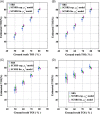
Functional Near-Infrared Spectroscopy (fNIRS) aims to recover changes in tissue optical parameters relating to tissue hemodynamics, to infer functional information in biological tissue. A widely-used application of fNIRS relies on continuous wave (CW) methodology that utilizes multiple distance measurements on human head for study of brain health. The typical method used is spatially resolved spectroscopy (SRS), which is shown to recover tissue oxygenation index (TOI) based on gradient of light intensity measured between two detectors. However, this methodology does not account for tissue scattering which is often assumed. A new parameter recovery algorithm is developed, which directly recovers both the scattering parameter and scaled chromophore concentrations and hence TOI from the measured gradient of light-attenuation at multiple wavelengths. It is shown through simulations that in comparison to conventional SRS which estimates cerebral TOI values with an error of ±12.3%, the proposed method provides more accurate estimate of TOI exhibiting an error of ±5.7% without any prior assumptions of tissue scatter, and can be easily implemented within CW fNIRS systems. Using an arm-cuff experiment, the obtained TOI using the proposed method is shown to provide a higher and more realistic value as compared to utilizing any prior assumptions of tissue scatter.
Keywords: near-infrared spectroscopy; tissue optics; tissue scattering.
© 2019 The Authors. Journal of Biophotonics published by WILEY-VCH Verlag GmbH & Co. KGaA, Weinheim.
Figures


References
-
- Stiefel M. F., Spiotta A., Gracias V. H., Garuffe A. M., Guillamondegui O., Maloney‐Wilensky E., Bloom S., Grady M. S., LeRoux P. D., J. Neurosurg. 2005, 103, 805. - PubMed
-
- Hodgkinson S., Pollit V., Sharpin C., Lecky F., BMJ 2014, 348, g104. - PubMed
-
- Maloney‐Wilensky E., Gracias V., Itkin A., Hoffman K., Bloom S., Yang W., Christian S., LeRoux P. D., Crit. Care Med. 2009, 37, 2057. - PubMed
-
- White H., Venkatesh B. in Oxford textbook of neurocritical care, chapter 17: traumatic brain injury, Vol., Oxford University Press, 2016, p. 213‐215.
-
- Torricelli A., Contini D., Pifferi A., Caffini M., Re R., Zucchelli L., Spinelli L., Neuroimage 2014, 85, 28. - PubMed
Publication types
MeSH terms
Substances
Grants and funding
LinkOut - more resources
Full Text Sources

