

At-line multi-angle light scattering detector for faster process development in enveloped virus-like particle purification
- PMID: 31169979
- PMCID: PMC6771681
- DOI: 10.1002/jssc.201900441
At-line multi-angle light scattering detector for faster process development in enveloped virus-like particle purification
Abstract
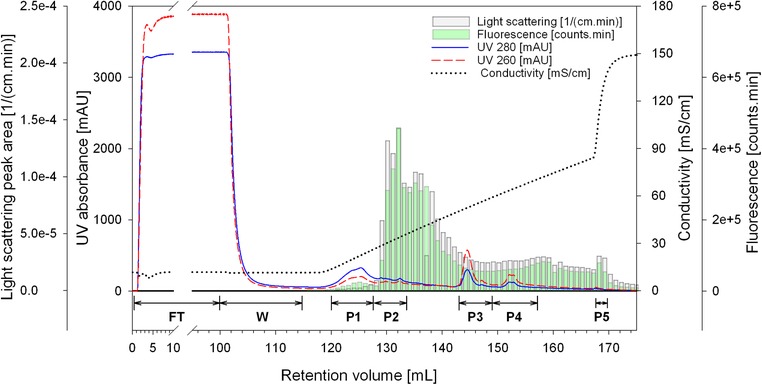
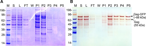
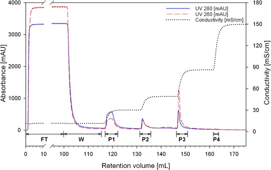
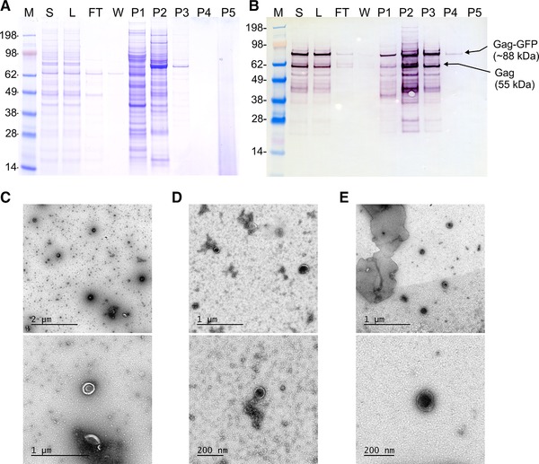
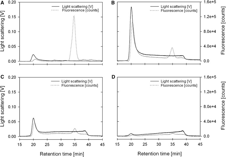
At-line static light scattering and fluorescence monitoring allows direct in-process tracking of fluorescent virus-like particles. We have demonstrated this by coupling at-line multi-angle light scattering and fluorescence detectors to the downstream processing of enveloped virus-like particles. Since light scattering intensity is directly proportional to particle concentration, our strategy allowed a swift identification of product containing fractions and rapid process development. Virus-like particles containing the Human Immunodeficiency Virus-1 Gag protein fused to the Green Fluorescence protein were produced in Human Embryonic Kidney 293 cells by transient transfection. A single-column anion-exchange chromatography method was used for direct capture and purification. The majority of host-cell protein impurities passed through the column without binding. Virus-like particles bound to the column were eluted by linear or step salt gradients. Particles recovered in the step gradient purification were characterized by nanoparticle tracking analysis, size exclusion chromatography coupled to multi-angle light scattering and fluorescence detectors and transmission electron microscopy. A total recovery of 66% for the fluorescent particles was obtained with a 50% yield in the main product peak. Virus-like particles were concentrated 17-fold to final a concentration of 4.45 × 10¹⁰ particles/mL. Simple buffers and operation make this process suitable for large scale purposes.
Keywords: enveloped bionanoparticles; fluorescent virus-like particles; monoliths; nanoparticle tracking analysis.
© 2019 The Authors. Journal of Separation Science published by WILEY-VCH Verlag GmbH & Co. KGaA, Weinheim.
Conflict of interest statement
The authors have declared no conflict of interest.
Figures
References
-
- Lua, L. H. , Connors, N. K. , Sainsbury, F. , Chuan, Y. P. , Wibowo, N. , Middelberg, A. P. , Bioengineering virus‐like particles as vaccines. Biotechnol. Bioeng. 2014, 111, 425–440. - PubMed
-
- Steppert, P. , Burgstaller, D. , Klausberger, M. , Tover, A. , Berger, E. , Jungbauer, A. , Quantification and characterization of virus‐like particles by size‐exclusion chromatography and nanoparticle tracking analysis. J. Chromatogr. A 2017, 1487, 89–99. - PubMed
-
- Bagheri, M. , Norouzi, H. R. , Hossienizadeh, S. M. J. , Es‐Haghi, A. , Ghassempour, A. , Development and modeling of two‐dimensional fast protein liquid chromatography for producing nonstructural protein‐free food‐and‐mouth diseases virus vaccine. J. Chromatogr. B 2018, 1096, 113–121. - PubMed
MeSH terms
Substances
Grants and funding
LinkOut - more resources
Full Text Sources
Other Literature Sources
