

Adenoviral gene transfer of a single-chain IL-23 induces psoriatic arthritis-like symptoms in NOD mice
- PMID: 31170010
- PMCID: PMC6662986
- DOI: 10.1096/fj.201900420R
Adenoviral gene transfer of a single-chain IL-23 induces psoriatic arthritis-like symptoms in NOD mice
Abstract
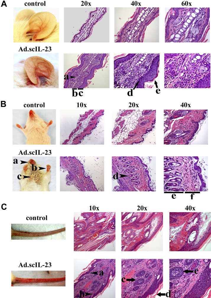
Previously, we demonstrated that intratumoral delivery of adenoviral vector encoding single-chain (sc)IL-23 (Ad.scIL-23) was able to induce systemic antitumor immunity. Here, we examined the role of IL-23 in diabetes in nonobese diabetic mice. Intravenous delivery of Ad.scIL-23 did not accelerate the onset of hyperglycemia but instead resulted in the development of psoriatic arthritis. Ad.scIL-23-treated mice developed erythema, scales, and thickening of the skin, as well as intervertebral disc degeneration and extensive synovial hypertrophy and loss of articular cartilage in the knees. Immunological analysis revealed activation of conventional T helper type 17 cells and IL-17-producing γδ T cells along with a significant depletion and suppression of T cells in the pancreatic lymph nodes. Furthermore, treatment with anti-IL-17 antibody reduced joint and skin psoriatic arthritis pathologies. Thus, these Ad.scIL-23-treated mice represent a physiologically relevant model of psoriatic arthritis for understanding disease progression and for testing therapeutic approaches.-Flores, R. R., Carbo, L., Kim, E., Van Meter, M., De Padilla, C. M. L., Zhao, J., Colangelo, D., Yousefzadeh, M. J., Angelini, L. A., Zhang, L., Pola, E., Vo, N., Evans, C. H., Gambotto, A., Niedernhofer, L. J., Robbins, P. D. Adenoviral gene transfer of a single-chain IL-23 induces psoriatic arthritis-like symptoms in NOD mice.
Keywords: adenovirus; interleukin-17; interleukin-23; psoriasis; type 1 diabetes.
Conflict of interest statement
The authors thank Biviana Torres (The Scripps Research Institute) for her assistance with flow cytometry and Shannon Howard, Sara McGowan, and Tokio Sano (all from The Scripps Research Institute) for their assistance in collecting and the processing of tissues. The authors acknowledge and extend gratitude to Yuan Yuan Ling (retired, formerly of The Scripps Research Institute) for years of service to science. This work was supported by Grants from the U.S. National Institutes of Health (NIH) National Institute on Aging (AG024827, AG044376), NIH National Institute of Arthritis and Musculoskeletal and Skin Diseases (AR051456), and a program grant from the Juvenile Diabetes Research Foundation to P.D.R. R.R.F. was supported by a T32 grant from NIH on Autoimmunity and Immunopathology. This project used the University of Pittsburgh Cancer Institute Vector Facilities (Pittsburgh, PA, USA) supported by the University of Pittsburgh’s NIH Cancer Center Support Grant P30 CA047904. The authors declare no conflicts of interest.
Figures


Similar articles
-
Differential Requirement for CCR6 in IL-23-Mediated Skin and Joint Inflammation.J Invest Dermatol. 2020 Dec;140(12):2386-2397. doi: 10.1016/j.jid.2020.03.965. Epub 2020 Apr 24. J Invest Dermatol. 2020. PMID: 32339538
-
IL-Y, a synthetic member of the IL-12 cytokine family, suppresses the development of type 1 diabetes in NOD mice.Eur J Immunol. 2015 Nov;45(11):3114-25. doi: 10.1002/eji.201445403. Epub 2015 Sep 1. Eur J Immunol. 2015. PMID: 26260044 Free PMC article.
-
Differential efficacy of biologic treatments targeting the TNF-α/IL-23/IL-17 axis in psoriasis and psoriatic arthritis.Cytokine. 2018 Nov;111:182-188. doi: 10.1016/j.cyto.2018.08.025. Epub 2018 Aug 29. Cytokine. 2018. PMID: 30172115 Review.
-
IL-23 induces CLEC5A+ IL-17A+ neutrophils and elicit skin inflammation associated with psoriatic arthritis.J Autoimmun. 2024 Feb;143:103167. doi: 10.1016/j.jaut.2024.103167. Epub 2024 Feb 1. J Autoimmun. 2024. PMID: 38301504 Free PMC article.
-
Role of the IL-23/IL-17 Axis in Psoriasis and Psoriatic Arthritis: The Clinical Importance of Its Divergence in Skin and Joints.Int J Mol Sci. 2018 Feb 9;19(2):530. doi: 10.3390/ijms19020530. Int J Mol Sci. 2018. PMID: 29425183 Free PMC article. Review.
Cited by
-
The Protective Role of pVHL in Imiquimod-Induced Psoriasis-like Skin Inflammation.Int J Mol Sci. 2022 May 7;23(9):5226. doi: 10.3390/ijms23095226. Int J Mol Sci. 2022. PMID: 35563616 Free PMC article.
-
Immune microenvironment in intervertebral disc degeneration: pathophysiology and therapeutic potential.Front Immunol. 2025 Jul 4;16:1563635. doi: 10.3389/fimmu.2025.1563635. eCollection 2025. Front Immunol. 2025. PMID: 40688090 Free PMC article. Review.
-
IL-23 orchestrating immune cell activation in arthritis.Rheumatology (Oxford). 2021 Oct 19;60(Suppl 4):iv4-iv15. doi: 10.1093/rheumatology/keab266. Rheumatology (Oxford). 2021. PMID: 34668017 Free PMC article.
-
What are mice teaching us about psoriatic arthritis?Curr Opin Rheumatol. 2025 Jul 1;37(4):243-253. doi: 10.1097/BOR.0000000000001093. Epub 2025 Apr 24. Curr Opin Rheumatol. 2025. PMID: 40265275 Free PMC article. Review.
-
Molecular Insights Into Lysyl Oxidases in Cartilage Regeneration and Rejuvenation.Front Bioeng Biotechnol. 2020 Apr 30;8:359. doi: 10.3389/fbioe.2020.00359. eCollection 2020. Front Bioeng Biotechnol. 2020. PMID: 32426343 Free PMC article. Review.
References
-
- Schön M. P., Boehncke W. H. (2005) Psoriasis. N. Engl. J. Med. 352, 1899–1912 - PubMed
-
- Baraliakos X., Coates L. C., Braun J. (2012) The involvement of the spine in psoriatic arthritis. J. Rheumatol. 39, 418–420 - PubMed
-
- Deng Y., Chang C., Lu Q. (2016) The inflammatory response in psoriasis: a comprehensive review. Clin. Rev. Allergy Immunol. 50, 377–389 - PubMed
Publication types
MeSH terms
Substances
Grants and funding
LinkOut - more resources
Full Text Sources
Medical
